Cobalt »
PDB 1a0c-1e1c »
1ao1 »
Cobalt in PDB 1ao1: Interactions of Deglycosylated Cobalt(III)-Pepleomycin with Dna, uc(Nmr), Minimized Average Structure
Cobalt Binding Sites:
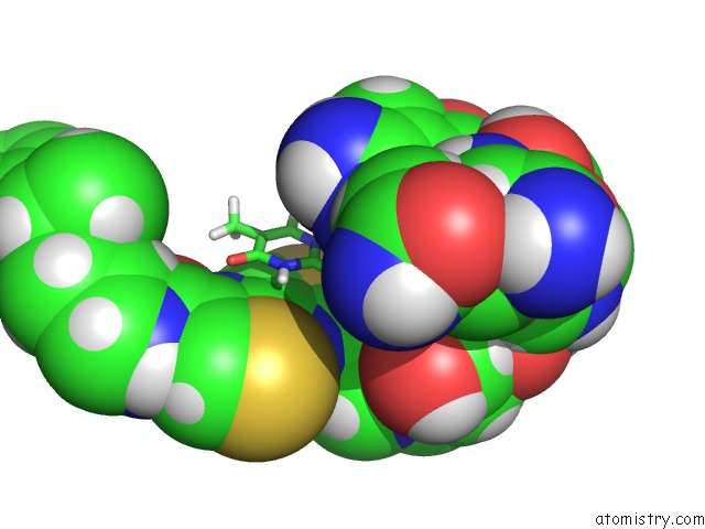
The binding sites of Cobalt atom in the Interactions of Deglycosylated Cobalt(III)-Pepleomycin with Dna, uc(Nmr), Minimized Average Structure
(pdb code 1ao1). This binding sites where shown within
5.0 Angstroms radius around Cobalt atom.
In total only one binding site of Cobalt was determined in the Interactions of Deglycosylated Cobalt(III)-Pepleomycin with Dna, uc(Nmr), Minimized Average Structure, PDB code: 1ao1:
In total only one binding site of Cobalt was determined in the Interactions of Deglycosylated Cobalt(III)-Pepleomycin with Dna, uc(Nmr), Minimized Average Structure, PDB code: 1ao1:
Cobalt binding site 1 out of 1 in 1ao1
Go back to
Cobalt binding site 1 out
of 1 in the Interactions of Deglycosylated Cobalt(III)-Pepleomycin with Dna, uc(Nmr), Minimized Average Structure
Mono view

Stereo pair view
Mono view
Stereo pair view
A full contact list of Cobalt with other atoms in the Co binding
site number 1 of Interactions of Deglycosylated Cobalt(III)-Pepleomycin with Dna, uc(Nmr), Minimized Average Structure within 5.0Å range:
|
Reference:
J.Caceres-Cortes,
H.Sugiyama,
K.Ikudome,
I.Saito,
A.H.Wang.
Interactions of Deglycosylated Cobalt(III)-Pepleomycin (Green Form) with Dna Based on uc(Nmr) Structural Studies,. Biochemistry V. 36 9995 1997.
ISSN: ISSN 0006-2960
PubMed: 9254594
DOI: 10.1021/BI9708951
Page generated: Sun Jul 13 17:22:27 2025
ISSN: ISSN 0006-2960
PubMed: 9254594
DOI: 10.1021/BI9708951
Last articles
Cu in 5IJUCu in 5ICU
Cu in 5I6P
Cu in 5IB3
Cu in 5I6O
Cu in 5I6N
Cu in 5I6M
Cu in 5I26
Cu in 5I6L
Cu in 5I38