Cobalt »
PDB 1a0c-1e1c »
1bmt »
Cobalt in PDB 1bmt: How A Protein Binds B12: A 3.O Angstrom X-Ray Structure of the B12-Binding Domains of Methionine Synthase
Enzymatic activity of How A Protein Binds B12: A 3.O Angstrom X-Ray Structure of the B12-Binding Domains of Methionine Synthase
All present enzymatic activity of How A Protein Binds B12: A 3.O Angstrom X-Ray Structure of the B12-Binding Domains of Methionine Synthase:
2.1.1.13;
2.1.1.13;
Protein crystallography data
The structure of How A Protein Binds B12: A 3.O Angstrom X-Ray Structure of the B12-Binding Domains of Methionine Synthase, PDB code: 1bmt
was solved by
C.L.Drennan,
S.Huang,
J.T.Drummond,
R.G.Matthews,
M.L.Ludwig,
with X-Ray Crystallography technique. A brief refinement statistics is given in the table below:
| Resolution Low / High (Å) | 8.00 / 3.00 |
| Space group | P 21 21 21 |
| Cell size a, b, c (Å), α, β, γ (°) | 96.700, 55.300, 103.800, 90.00, 90.00, 90.00 |
| R / Rfree (%) | 17 / n/a |
Cobalt Binding Sites:
The binding sites of Cobalt atom in the How A Protein Binds B12: A 3.O Angstrom X-Ray Structure of the B12-Binding Domains of Methionine Synthase
(pdb code 1bmt). This binding sites where shown within
5.0 Angstroms radius around Cobalt atom.
In total 2 binding sites of Cobalt where determined in the How A Protein Binds B12: A 3.O Angstrom X-Ray Structure of the B12-Binding Domains of Methionine Synthase, PDB code: 1bmt:
Jump to Cobalt binding site number: 1; 2;
In total 2 binding sites of Cobalt where determined in the How A Protein Binds B12: A 3.O Angstrom X-Ray Structure of the B12-Binding Domains of Methionine Synthase, PDB code: 1bmt:
Jump to Cobalt binding site number: 1; 2;
Cobalt binding site 1 out of 2 in 1bmt
Go back to
Cobalt binding site 1 out
of 2 in the How A Protein Binds B12: A 3.O Angstrom X-Ray Structure of the B12-Binding Domains of Methionine Synthase
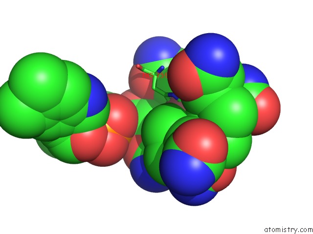
Mono view
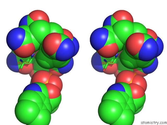
Stereo pair view
Mono view
Stereo pair view
A full contact list of Cobalt with other atoms in the Co binding
site number 1 of How A Protein Binds B12: A 3.O Angstrom X-Ray Structure of the B12-Binding Domains of Methionine Synthase within 5.0Å range:
|
Cobalt binding site 2 out of 2 in 1bmt
Go back to
Cobalt binding site 2 out
of 2 in the How A Protein Binds B12: A 3.O Angstrom X-Ray Structure of the B12-Binding Domains of Methionine Synthase
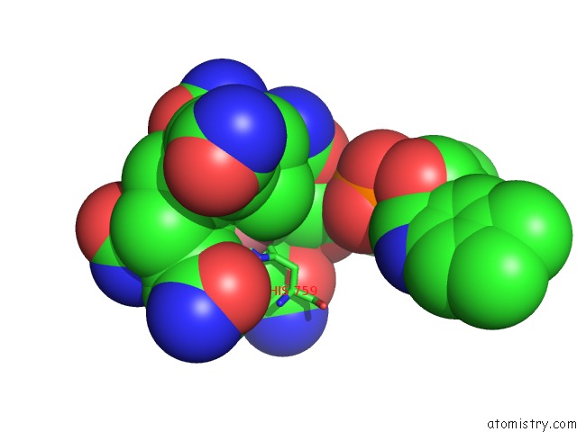
Mono view
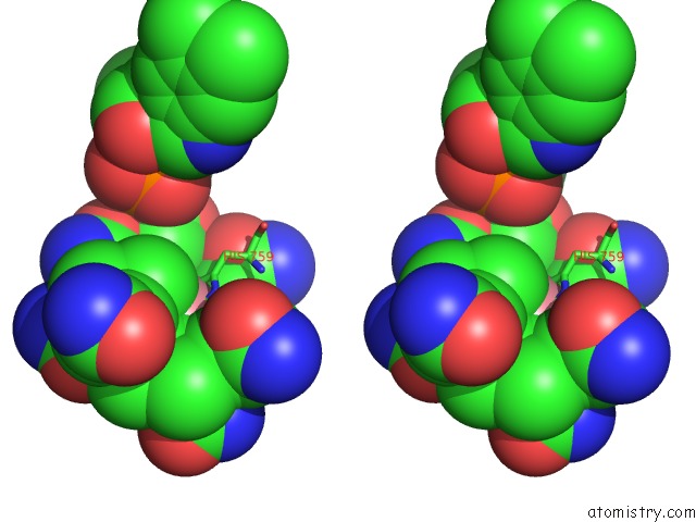
Stereo pair view
Mono view
Stereo pair view
A full contact list of Cobalt with other atoms in the Co binding
site number 2 of How A Protein Binds B12: A 3.O Angstrom X-Ray Structure of the B12-Binding Domains of Methionine Synthase within 5.0Å range:
|
Reference:
C.L.Drennan,
S.Huang,
J.T.Drummond,
R.G.Matthews,
M.L.Lidwig.
How A Protein Binds B12: A 3.0 A X-Ray Structure of B12-Binding Domains of Methionine Synthase. Science V. 266 1669 1994.
ISSN: ISSN 0036-8075
PubMed: 7992050
Page generated: Sun Jul 13 17:23:46 2025
ISSN: ISSN 0036-8075
PubMed: 7992050
Last articles
Cu in 3FYICu in 3FU9
Cu in 3FU8
Cu in 3FYE
Cu in 3FU7
Cu in 3FSW
Cu in 3FT0
Cu in 3FSZ
Cu in 3FPX
Cu in 3FSV