Cobalt »
PDB 1a0c-1e1c »
1boa »
Cobalt in PDB 1boa: Human Methionine Aminopeptidase 2 Complexed with Angiogenesis Inhibitor Fumagillin
Enzymatic activity of Human Methionine Aminopeptidase 2 Complexed with Angiogenesis Inhibitor Fumagillin
All present enzymatic activity of Human Methionine Aminopeptidase 2 Complexed with Angiogenesis Inhibitor Fumagillin:
3.4.11.18;
3.4.11.18;
Protein crystallography data
The structure of Human Methionine Aminopeptidase 2 Complexed with Angiogenesis Inhibitor Fumagillin, PDB code: 1boa
was solved by
S.Liu,
J.Widom,
C.W.Kemp,
C.M.Crews,
J.C.Clardy,
with X-Ray Crystallography technique. A brief refinement statistics is given in the table below:
| Resolution Low / High (Å) | 25.00 / 1.80 |
| Space group | C 2 2 21 |
| Cell size a, b, c (Å), α, β, γ (°) | 90.700, 99.580, 101.950, 90.00, 90.00, 90.00 |
| R / Rfree (%) | 19.3 / 23.2 |
Cobalt Binding Sites:
The binding sites of Cobalt atom in the Human Methionine Aminopeptidase 2 Complexed with Angiogenesis Inhibitor Fumagillin
(pdb code 1boa). This binding sites where shown within
5.0 Angstroms radius around Cobalt atom.
In total 2 binding sites of Cobalt where determined in the Human Methionine Aminopeptidase 2 Complexed with Angiogenesis Inhibitor Fumagillin, PDB code: 1boa:
Jump to Cobalt binding site number: 1; 2;
In total 2 binding sites of Cobalt where determined in the Human Methionine Aminopeptidase 2 Complexed with Angiogenesis Inhibitor Fumagillin, PDB code: 1boa:
Jump to Cobalt binding site number: 1; 2;
Cobalt binding site 1 out of 2 in 1boa
Go back to
Cobalt binding site 1 out
of 2 in the Human Methionine Aminopeptidase 2 Complexed with Angiogenesis Inhibitor Fumagillin
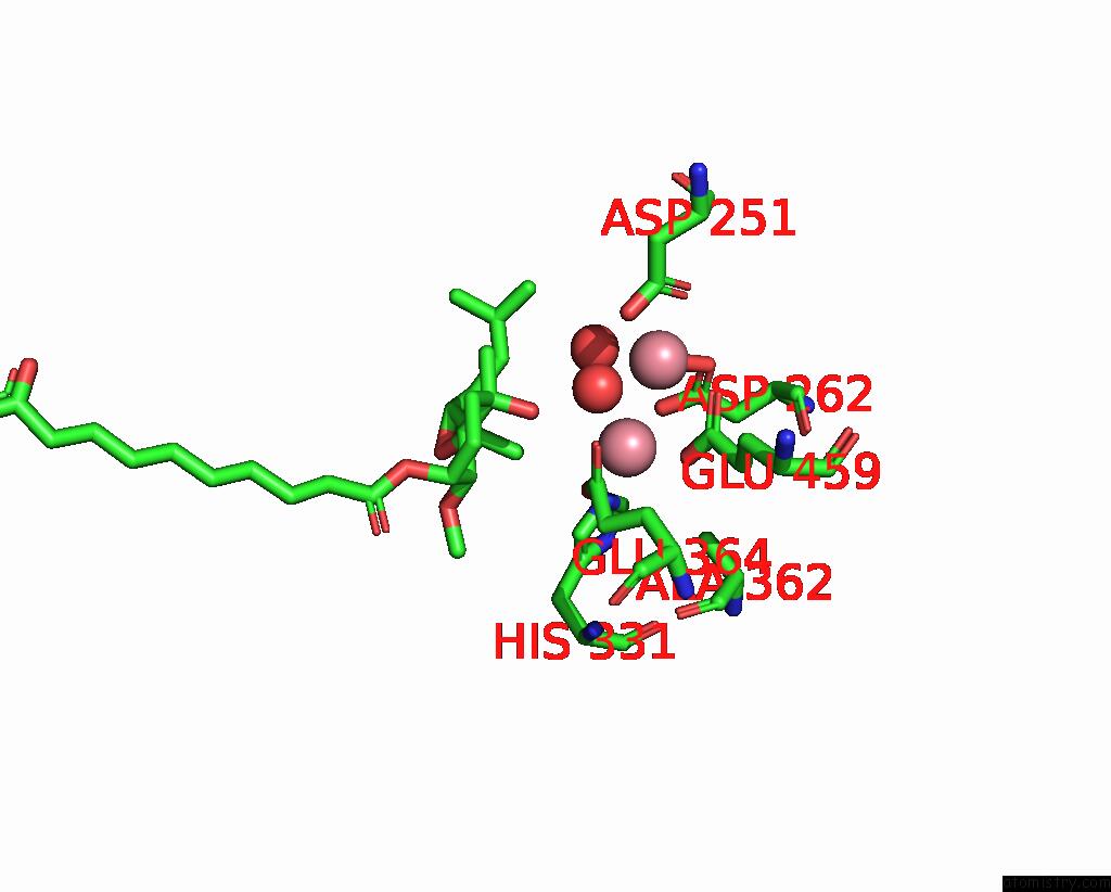
Mono view
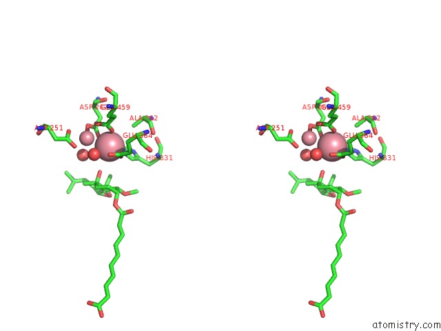
Stereo pair view
Mono view
Stereo pair view
A full contact list of Cobalt with other atoms in the Co binding
site number 1 of Human Methionine Aminopeptidase 2 Complexed with Angiogenesis Inhibitor Fumagillin within 5.0Å range:
|
Cobalt binding site 2 out of 2 in 1boa
Go back to
Cobalt binding site 2 out
of 2 in the Human Methionine Aminopeptidase 2 Complexed with Angiogenesis Inhibitor Fumagillin
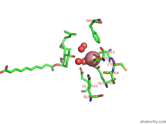
Mono view
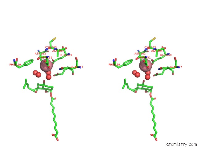
Stereo pair view
Mono view
Stereo pair view
A full contact list of Cobalt with other atoms in the Co binding
site number 2 of Human Methionine Aminopeptidase 2 Complexed with Angiogenesis Inhibitor Fumagillin within 5.0Å range:
|
Reference:
S.Liu,
J.Widom,
C.W.Kemp,
C.M.Crews,
J.Clardy.
Structure of Human Methionine Aminopeptidase-2 Complexed with Fumagillin. Science V. 282 1324 1998.
ISSN: ISSN 0036-8075
PubMed: 9812898
DOI: 10.1126/SCIENCE.282.5392.1324
Page generated: Sun Jul 13 17:24:02 2025
ISSN: ISSN 0036-8075
PubMed: 9812898
DOI: 10.1126/SCIENCE.282.5392.1324
Last articles
Cu in 5NQOCu in 5NQN
Cu in 5NQ9
Cu in 5O2X
Cu in 5O2W
Cu in 5NS5
Cu in 5NQM
Cu in 5NLT
Cu in 5NQ7
Cu in 5NQ8