Cobalt »
PDB 1a0c-1e1c »
1bsj »
Cobalt in PDB 1bsj: Cobalt Deformylase Inhibitor Complex From E.Coli
Enzymatic activity of Cobalt Deformylase Inhibitor Complex From E.Coli
All present enzymatic activity of Cobalt Deformylase Inhibitor Complex From E.Coli:
3.5.1.27;
3.5.1.27;
Protein crystallography data
The structure of Cobalt Deformylase Inhibitor Complex From E.Coli, PDB code: 1bsj
was solved by
B.Hao,
W.Gong,
P.T.Rajagopalan,
Y.Hu,
D.Pei,
M.K.Chan,
with X-Ray Crystallography technique. A brief refinement statistics is given in the table below:
| Resolution Low / High (Å) | 20.00 / 3.00 |
| Space group | P 65 2 2 |
| Cell size a, b, c (Å), α, β, γ (°) | 99.080, 99.080, 111.040, 90.00, 90.00, 120.00 |
| R / Rfree (%) | 16.8 / 20.7 |
Cobalt Binding Sites:
The binding sites of Cobalt atom in the Cobalt Deformylase Inhibitor Complex From E.Coli
(pdb code 1bsj). This binding sites where shown within
5.0 Angstroms radius around Cobalt atom.
In total only one binding site of Cobalt was determined in the Cobalt Deformylase Inhibitor Complex From E.Coli, PDB code: 1bsj:
In total only one binding site of Cobalt was determined in the Cobalt Deformylase Inhibitor Complex From E.Coli, PDB code: 1bsj:
Cobalt binding site 1 out of 1 in 1bsj
Go back to
Cobalt binding site 1 out
of 1 in the Cobalt Deformylase Inhibitor Complex From E.Coli
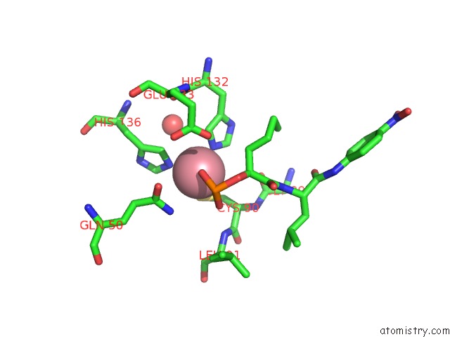
Mono view
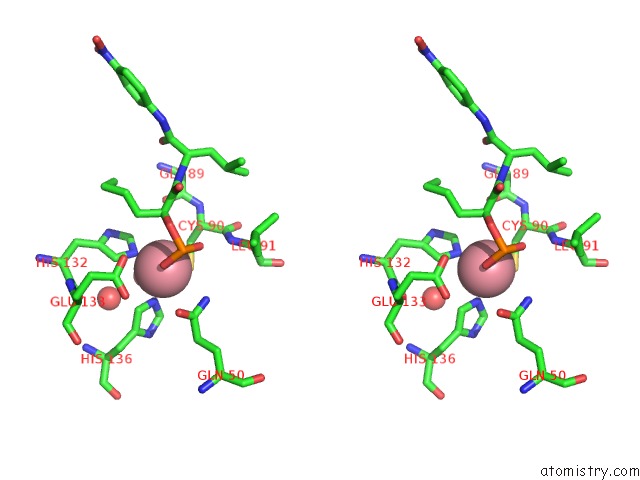
Stereo pair view
Mono view
Stereo pair view
A full contact list of Cobalt with other atoms in the Co binding
site number 1 of Cobalt Deformylase Inhibitor Complex From E.Coli within 5.0Å range:
|
Reference:
B.Hao,
W.Gong,
P.T.Rajagopalan,
Y.Zhou,
D.Pei,
M.K.Chan.
Structural Basis For the Design of Antibiotics Targeting Peptide Deformylase. Biochemistry V. 38 4712 1999.
ISSN: ISSN 0006-2960
PubMed: 10200158
DOI: 10.1021/BI982594C
Page generated: Sun Jul 13 17:24:13 2025
ISSN: ISSN 0006-2960
PubMed: 10200158
DOI: 10.1021/BI982594C
Last articles
Cu in 4E9XCu in 4E9Y
Cu in 4E9V
Cu in 4E9W
Cu in 4BED
Cu in 4E9T
Cu in 4E9R
Cu in 4E9S
Cu in 4E9Q
Cu in 4E4Z