Cobalt »
PDB 1a0c-1e1c »
1diy »
Cobalt in PDB 1diy: Crystal Structure of Arachidonic Acid Bound in the Cyclooxygenase Active Site of Pghs-1
Enzymatic activity of Crystal Structure of Arachidonic Acid Bound in the Cyclooxygenase Active Site of Pghs-1
All present enzymatic activity of Crystal Structure of Arachidonic Acid Bound in the Cyclooxygenase Active Site of Pghs-1:
1.14.99.1;
1.14.99.1;
Protein crystallography data
The structure of Crystal Structure of Arachidonic Acid Bound in the Cyclooxygenase Active Site of Pghs-1, PDB code: 1diy
was solved by
M.G.Malkowski,
S.L.Ginell,
W.L.Smith,
R.M.Garavito,
with X-Ray Crystallography technique. A brief refinement statistics is given in the table below:
| Resolution Low / High (Å) | 9.00 / 3.00 |
| Space group | P 65 2 2 |
| Cell size a, b, c (Å), α, β, γ (°) | 182.100, 182.100, 103.640, 90.00, 90.00, 120.00 |
| R / Rfree (%) | 21.5 / 29 |
Cobalt Binding Sites:
The binding sites of Cobalt atom in the Crystal Structure of Arachidonic Acid Bound in the Cyclooxygenase Active Site of Pghs-1
(pdb code 1diy). This binding sites where shown within
5.0 Angstroms radius around Cobalt atom.
In total only one binding site of Cobalt was determined in the Crystal Structure of Arachidonic Acid Bound in the Cyclooxygenase Active Site of Pghs-1, PDB code: 1diy:
In total only one binding site of Cobalt was determined in the Crystal Structure of Arachidonic Acid Bound in the Cyclooxygenase Active Site of Pghs-1, PDB code: 1diy:
Cobalt binding site 1 out of 1 in 1diy
Go back to
Cobalt binding site 1 out
of 1 in the Crystal Structure of Arachidonic Acid Bound in the Cyclooxygenase Active Site of Pghs-1
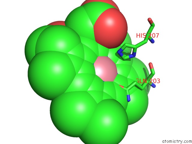
Mono view
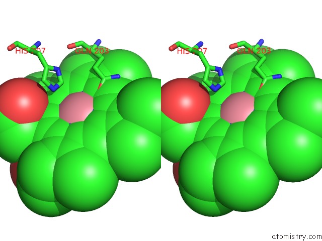
Stereo pair view
Mono view
Stereo pair view
A full contact list of Cobalt with other atoms in the Co binding
site number 1 of Crystal Structure of Arachidonic Acid Bound in the Cyclooxygenase Active Site of Pghs-1 within 5.0Å range:
|
Reference:
M.G.Malkowski,
S.L.Ginell,
W.L.Smith,
R.M.Garavito.
The Productive Conformation of Arachidonic Acid Bound to Prostaglandin Synthase. Science V. 289 1933 2000.
ISSN: ISSN 0036-8075
PubMed: 10988074
DOI: 10.1126/SCIENCE.289.5486.1933
Page generated: Sun Jul 13 17:26:47 2025
ISSN: ISSN 0036-8075
PubMed: 10988074
DOI: 10.1126/SCIENCE.289.5486.1933
Last articles
Cu in 4P5RCu in 4OUA
Cu in 4OZ7
Cu in 4NQQ
Cu in 4OY6
Cu in 4OPB
Cu in 4OJA
Cu in 4OAK
Cu in 4N8U
Cu in 4O65