Cobalt »
PDB 1hv9-1nr5 »
1ire »
Cobalt in PDB 1ire: Crystal Structure of Co-Type Nitrile Hydratase From Pseudonocardia Thermophila
Enzymatic activity of Crystal Structure of Co-Type Nitrile Hydratase From Pseudonocardia Thermophila
All present enzymatic activity of Crystal Structure of Co-Type Nitrile Hydratase From Pseudonocardia Thermophila:
4.2.1.84;
4.2.1.84;
Protein crystallography data
The structure of Crystal Structure of Co-Type Nitrile Hydratase From Pseudonocardia Thermophila, PDB code: 1ire
was solved by
A.Miyanaga,
S.Fushinobu,
K.Ito,
T.Wakagi,
with X-Ray Crystallography technique. A brief refinement statistics is given in the table below:
| Resolution Low / High (Å) | 28.03 / 1.80 |
| Space group | P 32 2 1 |
| Cell size a, b, c (Å), α, β, γ (°) | 65.501, 65.501, 184.614, 90.00, 90.00, 120.00 |
| R / Rfree (%) | 17.8 / 19.1 |
Cobalt Binding Sites:
The binding sites of Cobalt atom in the Crystal Structure of Co-Type Nitrile Hydratase From Pseudonocardia Thermophila
(pdb code 1ire). This binding sites where shown within
5.0 Angstroms radius around Cobalt atom.
In total only one binding site of Cobalt was determined in the Crystal Structure of Co-Type Nitrile Hydratase From Pseudonocardia Thermophila, PDB code: 1ire:
In total only one binding site of Cobalt was determined in the Crystal Structure of Co-Type Nitrile Hydratase From Pseudonocardia Thermophila, PDB code: 1ire:
Cobalt binding site 1 out of 1 in 1ire
Go back to
Cobalt binding site 1 out
of 1 in the Crystal Structure of Co-Type Nitrile Hydratase From Pseudonocardia Thermophila
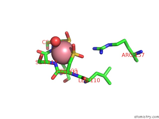
Mono view
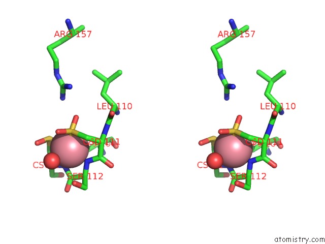
Stereo pair view
Mono view
Stereo pair view
A full contact list of Cobalt with other atoms in the Co binding
site number 1 of Crystal Structure of Co-Type Nitrile Hydratase From Pseudonocardia Thermophila within 5.0Å range:
|
Reference:
A.Miyanaga,
S.Fushinobu,
K.Ito,
T.Wakagi.
Crystal Structure of Cobalt-Containing Nitrile Hydratase. Biochem.Biophys.Res.Commun. V. 288 1169 2001.
ISSN: ISSN 0006-291X
PubMed: 11700034
DOI: 10.1006/BBRC.2001.5897
Page generated: Tue Jul 30 14:23:23 2024
ISSN: ISSN 0006-291X
PubMed: 11700034
DOI: 10.1006/BBRC.2001.5897
Last articles
Zn in 9J0NZn in 9J0O
Zn in 9J0P
Zn in 9FJX
Zn in 9EKB
Zn in 9C0F
Zn in 9CAH
Zn in 9CH0
Zn in 9CH3
Zn in 9CH1