Cobalt »
PDB 1hv9-1nr5 »
1jn1 »
Cobalt in PDB 1jn1: Structure of 2C-Methyl-D-Erythritol 2,4-Cyclodiphosphate Synthase From Haemophilus Influenzae (HI0671)
Protein crystallography data
The structure of Structure of 2C-Methyl-D-Erythritol 2,4-Cyclodiphosphate Synthase From Haemophilus Influenzae (HI0671), PDB code: 1jn1
was solved by
C.Lehmann,
K.Lim,
J.Toedt,
W.Krajewski,
A.Howard,
E.Eisenstein,
O.Herzberg,
Structure 2 Function Project (S2F),
with X-Ray Crystallography technique. A brief refinement statistics is given in the table below:
| Resolution Low / High (Å) | 20.00 / 2.90 |
| Space group | I 41 2 2 |
| Cell size a, b, c (Å), α, β, γ (°) | 112.300, 112.300, 187.600, 90.00, 90.00, 90.00 |
| R / Rfree (%) | 19.8 / 29 |
Cobalt Binding Sites:
The binding sites of Cobalt atom in the Structure of 2C-Methyl-D-Erythritol 2,4-Cyclodiphosphate Synthase From Haemophilus Influenzae (HI0671)
(pdb code 1jn1). This binding sites where shown within
5.0 Angstroms radius around Cobalt atom.
In total 4 binding sites of Cobalt where determined in the Structure of 2C-Methyl-D-Erythritol 2,4-Cyclodiphosphate Synthase From Haemophilus Influenzae (HI0671), PDB code: 1jn1:
Jump to Cobalt binding site number: 1; 2; 3; 4;
In total 4 binding sites of Cobalt where determined in the Structure of 2C-Methyl-D-Erythritol 2,4-Cyclodiphosphate Synthase From Haemophilus Influenzae (HI0671), PDB code: 1jn1:
Jump to Cobalt binding site number: 1; 2; 3; 4;
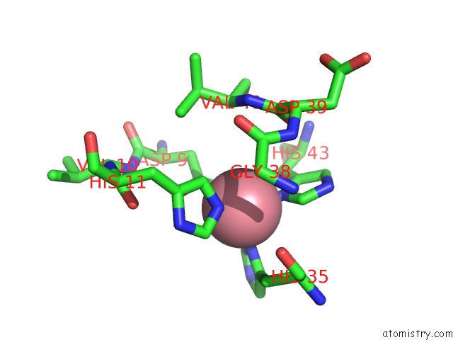
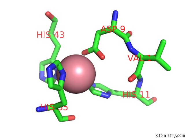
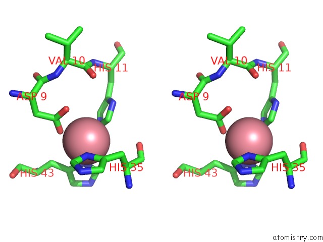
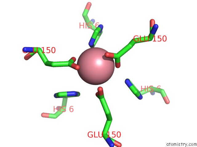
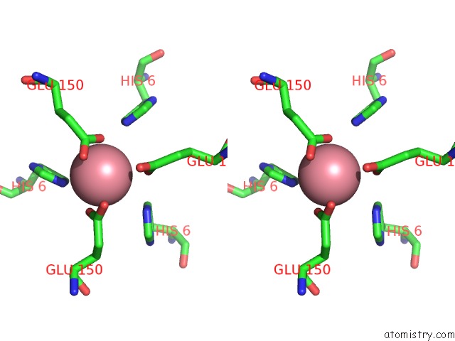
Cobalt binding site 1 out of 4 in 1jn1
Go back to
Cobalt binding site 1 out
of 4 in the Structure of 2C-Methyl-D-Erythritol 2,4-Cyclodiphosphate Synthase From Haemophilus Influenzae (HI0671)
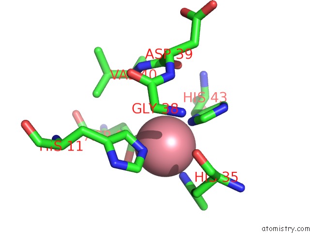
Mono view

Stereo pair view
Mono view
Stereo pair view
A full contact list of Cobalt with other atoms in the Co binding
site number 1 of Structure of 2C-Methyl-D-Erythritol 2,4-Cyclodiphosphate Synthase From Haemophilus Influenzae (HI0671) within 5.0Å range:
|
Cobalt binding site 2 out of 4 in 1jn1
Go back to
Cobalt binding site 2 out
of 4 in the Structure of 2C-Methyl-D-Erythritol 2,4-Cyclodiphosphate Synthase From Haemophilus Influenzae (HI0671)
Mono view

Stereo pair view
Mono view
Stereo pair view
A full contact list of Cobalt with other atoms in the Co binding
site number 2 of Structure of 2C-Methyl-D-Erythritol 2,4-Cyclodiphosphate Synthase From Haemophilus Influenzae (HI0671) within 5.0Å range:
|
Cobalt binding site 3 out of 4 in 1jn1
Go back to
Cobalt binding site 3 out
of 4 in the Structure of 2C-Methyl-D-Erythritol 2,4-Cyclodiphosphate Synthase From Haemophilus Influenzae (HI0671)
Mono view
Stereo pair view
Mono view
Stereo pair view
A full contact list of Cobalt with other atoms in the Co binding
site number 3 of Structure of 2C-Methyl-D-Erythritol 2,4-Cyclodiphosphate Synthase From Haemophilus Influenzae (HI0671) within 5.0Å range:
|
Cobalt binding site 4 out of 4 in 1jn1
Go back to
Cobalt binding site 4 out
of 4 in the Structure of 2C-Methyl-D-Erythritol 2,4-Cyclodiphosphate Synthase From Haemophilus Influenzae (HI0671)
Mono view
Stereo pair view
Mono view
Stereo pair view
A full contact list of Cobalt with other atoms in the Co binding
site number 4 of Structure of 2C-Methyl-D-Erythritol 2,4-Cyclodiphosphate Synthase From Haemophilus Influenzae (HI0671) within 5.0Å range:
|
Reference:
C.Lehmann,
K.Lim,
J.Toedt,
W.Krajewski,
A.Howard,
E.Eisenstein,
O.Herzberg.
Structure of 2C-Methyl-D-Erythrol-2,4-Cyclodiphosphate Synthase From Haemophilus Influenzae: Activation By Conformational Transition. Proteins V. 49 135 2002.
ISSN: ISSN 0887-3585
PubMed: 12211023
DOI: 10.1002/PROT.10182
Page generated: Tue Jul 30 14:23:31 2024
ISSN: ISSN 0887-3585
PubMed: 12211023
DOI: 10.1002/PROT.10182
Last articles
Zn in 9J0NZn in 9J0O
Zn in 9J0P
Zn in 9FJX
Zn in 9EKB
Zn in 9C0F
Zn in 9CAH
Zn in 9CH0
Zn in 9CH3
Zn in 9CH1