Cobalt »
PDB 1hv9-1nr5 »
1n4a »
Cobalt in PDB 1n4a: The Ligand Bound Structure of E.Coli Btuf, the Periplasmic Binding Protein For Vitamin B12
Protein crystallography data
The structure of The Ligand Bound Structure of E.Coli Btuf, the Periplasmic Binding Protein For Vitamin B12, PDB code: 1n4a
was solved by
N.K.Karpowich,
P.C.Smith,
J.F.Hunt,
Northeast Structural Genomicsconsortium (Nesg),
with X-Ray Crystallography technique. A brief refinement statistics is given in the table below:
| Resolution Low / High (Å) | 20.00 / 2.00 |
| Space group | P 21 21 2 |
| Cell size a, b, c (Å), α, β, γ (°) | 131.730, 92.020, 44.690, 90.00, 90.00, 90.00 |
| R / Rfree (%) | 23.4 / 26.1 |
Cobalt Binding Sites:
The binding sites of Cobalt atom in the The Ligand Bound Structure of E.Coli Btuf, the Periplasmic Binding Protein For Vitamin B12
(pdb code 1n4a). This binding sites where shown within
5.0 Angstroms radius around Cobalt atom.
In total 2 binding sites of Cobalt where determined in the The Ligand Bound Structure of E.Coli Btuf, the Periplasmic Binding Protein For Vitamin B12, PDB code: 1n4a:
Jump to Cobalt binding site number: 1; 2;
In total 2 binding sites of Cobalt where determined in the The Ligand Bound Structure of E.Coli Btuf, the Periplasmic Binding Protein For Vitamin B12, PDB code: 1n4a:
Jump to Cobalt binding site number: 1; 2;
Cobalt binding site 1 out of 2 in 1n4a
Go back to
Cobalt binding site 1 out
of 2 in the The Ligand Bound Structure of E.Coli Btuf, the Periplasmic Binding Protein For Vitamin B12
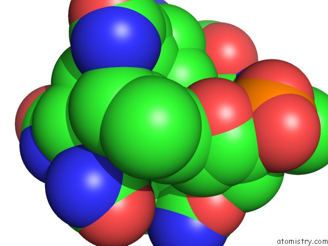
Mono view
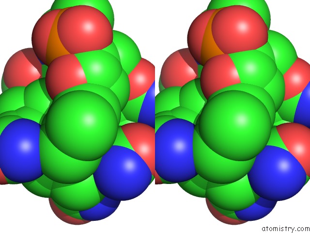
Stereo pair view
Mono view
Stereo pair view
A full contact list of Cobalt with other atoms in the Co binding
site number 1 of The Ligand Bound Structure of E.Coli Btuf, the Periplasmic Binding Protein For Vitamin B12 within 5.0Å range:
|
Cobalt binding site 2 out of 2 in 1n4a
Go back to
Cobalt binding site 2 out
of 2 in the The Ligand Bound Structure of E.Coli Btuf, the Periplasmic Binding Protein For Vitamin B12
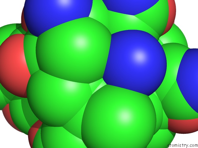
Mono view
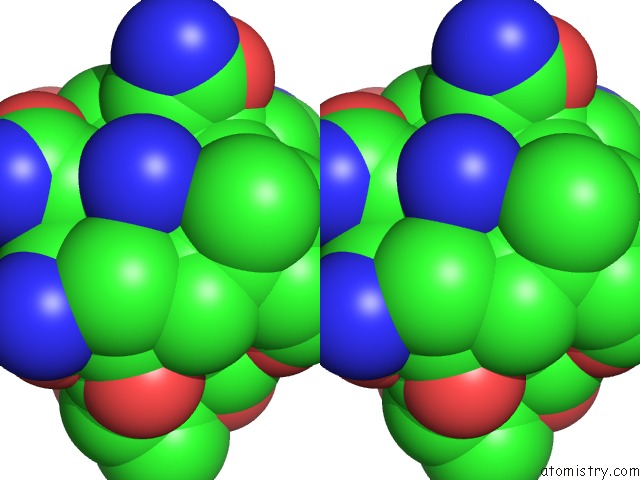
Stereo pair view
Mono view
Stereo pair view
A full contact list of Cobalt with other atoms in the Co binding
site number 2 of The Ligand Bound Structure of E.Coli Btuf, the Periplasmic Binding Protein For Vitamin B12 within 5.0Å range:
|
Reference:
N.K.Karpowich,
H.H.Huang,
P.C.Smith,
J.F.Hunt.
Crystal Structures of the Btuf Periplasmic-Binding Protein For Vitamin B12 Suggest A Functionally Important Reduction in Protein Mobility Upon Ligand Binding. J.Biol.Chem. V. 278 8429 2003.
ISSN: ISSN 0021-9258
PubMed: 12468528
DOI: 10.1074/JBC.M212239200
Page generated: Tue Jul 30 14:27:14 2024
ISSN: ISSN 0021-9258
PubMed: 12468528
DOI: 10.1074/JBC.M212239200
Last articles
Zn in 9MJ5Zn in 9HNW
Zn in 9G0L
Zn in 9FNE
Zn in 9DZN
Zn in 9E0I
Zn in 9D32
Zn in 9DAK
Zn in 8ZXC
Zn in 8ZUF