Cobalt »
PDB 1nyi-1rr2 »
1qk0 »
Cobalt in PDB 1qk0: CEL6A with A Non-Hydrolysable Cellotetraose
Enzymatic activity of CEL6A with A Non-Hydrolysable Cellotetraose
All present enzymatic activity of CEL6A with A Non-Hydrolysable Cellotetraose:
3.2.1.91;
3.2.1.91;
Protein crystallography data
The structure of CEL6A with A Non-Hydrolysable Cellotetraose, PDB code: 1qk0
was solved by
J.-Y.Zou,
T.A.Jones,
with X-Ray Crystallography technique. A brief refinement statistics is given in the table below:
| Resolution Low / High (Å) | 20.00 / 2.10 |
| Space group | P 1 21 1 |
| Cell size a, b, c (Å), α, β, γ (°) | 48.500, 74.690, 91.140, 90.00, 103.19, 90.00 |
| R / Rfree (%) | 18.1 / 22.1 |
Other elements in 1qk0:
The structure of CEL6A with A Non-Hydrolysable Cellotetraose also contains other interesting chemical elements:
| Iodine | (I) | 3 atoms |
Cobalt Binding Sites:
The binding sites of Cobalt atom in the CEL6A with A Non-Hydrolysable Cellotetraose
(pdb code 1qk0). This binding sites where shown within
5.0 Angstroms radius around Cobalt atom.
In total 2 binding sites of Cobalt where determined in the CEL6A with A Non-Hydrolysable Cellotetraose, PDB code: 1qk0:
Jump to Cobalt binding site number: 1; 2;
In total 2 binding sites of Cobalt where determined in the CEL6A with A Non-Hydrolysable Cellotetraose, PDB code: 1qk0:
Jump to Cobalt binding site number: 1; 2;
Cobalt binding site 1 out of 2 in 1qk0
Go back to
Cobalt binding site 1 out
of 2 in the CEL6A with A Non-Hydrolysable Cellotetraose
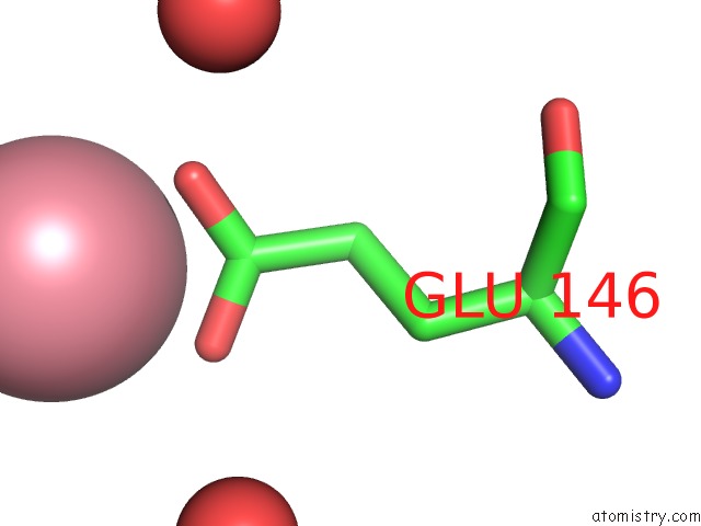
Mono view
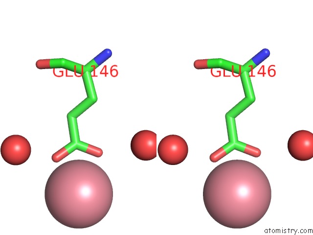
Stereo pair view
Mono view
Stereo pair view
A full contact list of Cobalt with other atoms in the Co binding
site number 1 of CEL6A with A Non-Hydrolysable Cellotetraose within 5.0Å range:
|
Cobalt binding site 2 out of 2 in 1qk0
Go back to
Cobalt binding site 2 out
of 2 in the CEL6A with A Non-Hydrolysable Cellotetraose
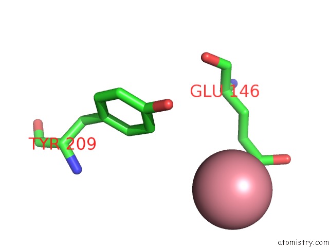
Mono view
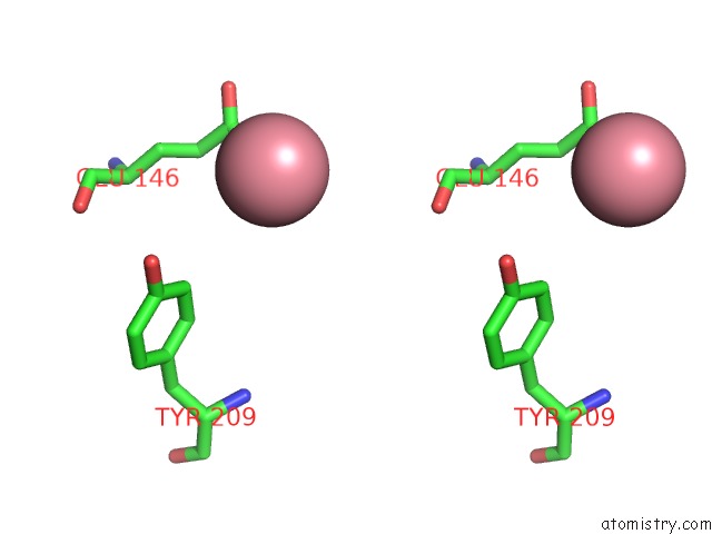
Stereo pair view
Mono view
Stereo pair view
A full contact list of Cobalt with other atoms in the Co binding
site number 2 of CEL6A with A Non-Hydrolysable Cellotetraose within 5.0Å range:
|
Reference:
J.-Y.Zou,
G.J.Kleywegt,
J.Stahlberg,
H.Driguez,
W.Nerinckx,
M.Claeyssens,
A.Koivula,
T.T.Teeri,
T.A.Jones.
Crystallographic Evidence For Substrate Ring Distortion and Protein Conformational Changes During Catalysis in Cellobiohydrolase CEL6A From Trichoderma Reesei Structure V. 7 1035 1999.
ISSN: ISSN 0969-2126
PubMed: 10508787
DOI: 10.1016/S0969-2126(99)80171-3
Page generated: Tue Jul 30 14:31:12 2024
ISSN: ISSN 0969-2126
PubMed: 10508787
DOI: 10.1016/S0969-2126(99)80171-3
Last articles
Zn in 9JYWZn in 9IR4
Zn in 9IR3
Zn in 9GMX
Zn in 9GMW
Zn in 9JEJ
Zn in 9ERF
Zn in 9ERE
Zn in 9EGV
Zn in 9EGW