Cobalt »
PDB 1nyi-1rr2 »
1qxz »
Cobalt in PDB 1qxz: Crystal Structure of S. Aureus Methionine Aminopeptidase in Complex with A Ketoheterocycle Inhibitor 119
Enzymatic activity of Crystal Structure of S. Aureus Methionine Aminopeptidase in Complex with A Ketoheterocycle Inhibitor 119
All present enzymatic activity of Crystal Structure of S. Aureus Methionine Aminopeptidase in Complex with A Ketoheterocycle Inhibitor 119:
3.4.11.18;
3.4.11.18;
Protein crystallography data
The structure of Crystal Structure of S. Aureus Methionine Aminopeptidase in Complex with A Ketoheterocycle Inhibitor 119, PDB code: 1qxz
was solved by
A.Douangamath,
G.E.Dale,
A.D'arcy,
C.Oefner,
with X-Ray Crystallography technique. A brief refinement statistics is given in the table below:
| Resolution Low / High (Å) | 20.00 / 1.68 |
| Space group | P 1 21 1 |
| Cell size a, b, c (Å), α, β, γ (°) | 40.947, 76.916, 41.651, 90.00, 104.79, 90.00 |
| R / Rfree (%) | 17 / 19.9 |
Cobalt Binding Sites:
The binding sites of Cobalt atom in the Crystal Structure of S. Aureus Methionine Aminopeptidase in Complex with A Ketoheterocycle Inhibitor 119
(pdb code 1qxz). This binding sites where shown within
5.0 Angstroms radius around Cobalt atom.
In total 3 binding sites of Cobalt where determined in the Crystal Structure of S. Aureus Methionine Aminopeptidase in Complex with A Ketoheterocycle Inhibitor 119, PDB code: 1qxz:
Jump to Cobalt binding site number: 1; 2; 3;
In total 3 binding sites of Cobalt where determined in the Crystal Structure of S. Aureus Methionine Aminopeptidase in Complex with A Ketoheterocycle Inhibitor 119, PDB code: 1qxz:
Jump to Cobalt binding site number: 1; 2; 3;
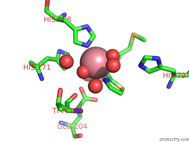
Cobalt binding site 1 out of 3 in 1qxz
Go back to
Cobalt binding site 1 out
of 3 in the Crystal Structure of S. Aureus Methionine Aminopeptidase in Complex with A Ketoheterocycle Inhibitor 119
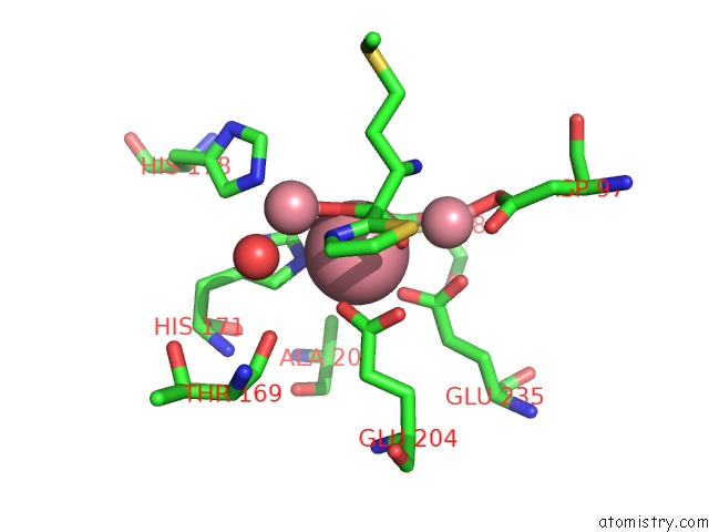
Mono view
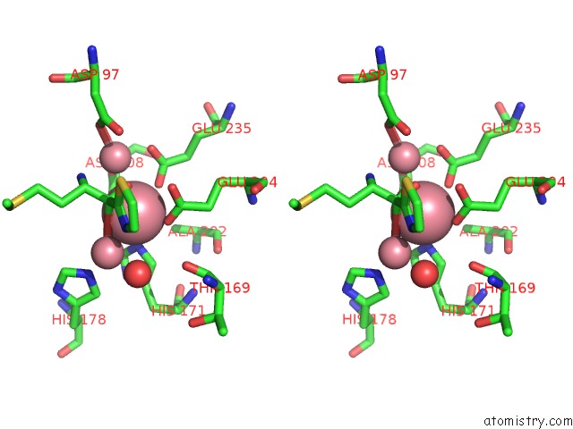
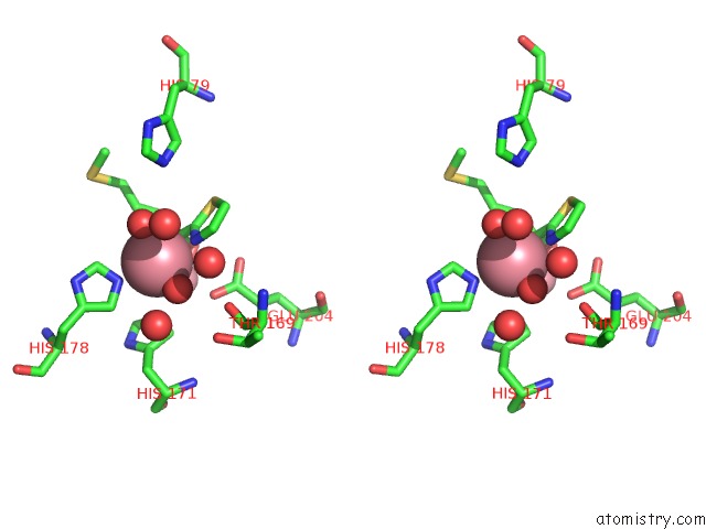
Stereo pair view
Mono view
Stereo pair view
A full contact list of Cobalt with other atoms in the Co binding
site number 1 of Crystal Structure of S. Aureus Methionine Aminopeptidase in Complex with A Ketoheterocycle Inhibitor 119 within 5.0Å range:
|
Cobalt binding site 2 out of 3 in 1qxz
Go back to
Cobalt binding site 2 out
of 3 in the Crystal Structure of S. Aureus Methionine Aminopeptidase in Complex with A Ketoheterocycle Inhibitor 119
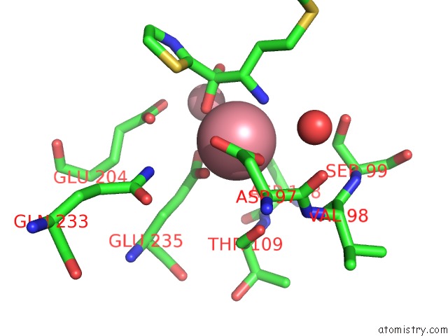
Mono view
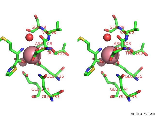
Stereo pair view
Mono view
Stereo pair view
A full contact list of Cobalt with other atoms in the Co binding
site number 2 of Crystal Structure of S. Aureus Methionine Aminopeptidase in Complex with A Ketoheterocycle Inhibitor 119 within 5.0Å range:
|
Cobalt binding site 3 out of 3 in 1qxz
Go back to
Cobalt binding site 3 out
of 3 in the Crystal Structure of S. Aureus Methionine Aminopeptidase in Complex with A Ketoheterocycle Inhibitor 119
Mono view
Stereo pair view
Mono view
Stereo pair view
A full contact list of Cobalt with other atoms in the Co binding
site number 3 of Crystal Structure of S. Aureus Methionine Aminopeptidase in Complex with A Ketoheterocycle Inhibitor 119 within 5.0Å range:
|
Reference:
A.Douangamath,
G.E.Dale,
A.D'arcy,
M.Almstetter,
R.Eckl,
A.Frutos-Hoener,
B.Henkel,
K.Illgen,
S.Nerdinger,
H.Schulz,
A.Macsweeney,
M.Thormann,
A.Treml,
S.Pierau,
S.Wadman,
C.Oefner.
Crystal Structures of Staphylococcusaureus Methionine Aminopeptidase Complexed with Keto Heterocycle and Aminoketone Inhibitors Reveal the Formation of A Tetrahedral Intermediate. J.Med.Chem. V. 47 1325 2004.
ISSN: ISSN 0022-2623
PubMed: 14998322
DOI: 10.1021/JM034188J
Page generated: Tue Jul 30 14:32:58 2024
ISSN: ISSN 0022-2623
PubMed: 14998322
DOI: 10.1021/JM034188J
Last articles
Zn in 9J0NZn in 9J0O
Zn in 9J0P
Zn in 9FJX
Zn in 9EKB
Zn in 9C0F
Zn in 9CAH
Zn in 9CH0
Zn in 9CH3
Zn in 9CH1