Cobalt »
PDB 1rrk-1vlx »
1u67 »
Cobalt in PDB 1u67: Crystal Structure of Arachidonic Acid Bound to A Mutant of Prostagladin H Synthase-1 That Forms Predominantly 11-Hpete.
Enzymatic activity of Crystal Structure of Arachidonic Acid Bound to A Mutant of Prostagladin H Synthase-1 That Forms Predominantly 11-Hpete.
All present enzymatic activity of Crystal Structure of Arachidonic Acid Bound to A Mutant of Prostagladin H Synthase-1 That Forms Predominantly 11-Hpete.:
1.14.99.1;
1.14.99.1;
Protein crystallography data
The structure of Crystal Structure of Arachidonic Acid Bound to A Mutant of Prostagladin H Synthase-1 That Forms Predominantly 11-Hpete., PDB code: 1u67
was solved by
C.A.Harman,
C.J.Rieke,
R.M.Garavito,
W.L.Smith,
with X-Ray Crystallography technique. A brief refinement statistics is given in the table below:
| Resolution Low / High (Å) | 20.00 / 3.10 |
| Space group | P 65 2 2 |
| Cell size a, b, c (Å), α, β, γ (°) | 181.879, 181.879, 103.368, 90.00, 90.00, 120.00 |
| R / Rfree (%) | 23.8 / 31.1 |
Cobalt Binding Sites:
The binding sites of Cobalt atom in the Crystal Structure of Arachidonic Acid Bound to A Mutant of Prostagladin H Synthase-1 That Forms Predominantly 11-Hpete.
(pdb code 1u67). This binding sites where shown within
5.0 Angstroms radius around Cobalt atom.
In total only one binding site of Cobalt was determined in the Crystal Structure of Arachidonic Acid Bound to A Mutant of Prostagladin H Synthase-1 That Forms Predominantly 11-Hpete., PDB code: 1u67:
In total only one binding site of Cobalt was determined in the Crystal Structure of Arachidonic Acid Bound to A Mutant of Prostagladin H Synthase-1 That Forms Predominantly 11-Hpete., PDB code: 1u67:
Cobalt binding site 1 out of 1 in 1u67
Go back to
Cobalt binding site 1 out
of 1 in the Crystal Structure of Arachidonic Acid Bound to A Mutant of Prostagladin H Synthase-1 That Forms Predominantly 11-Hpete.
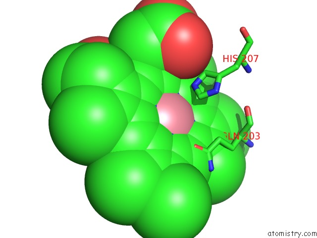
Mono view
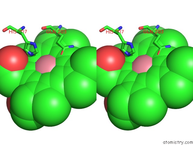
Stereo pair view
Mono view
Stereo pair view
A full contact list of Cobalt with other atoms in the Co binding
site number 1 of Crystal Structure of Arachidonic Acid Bound to A Mutant of Prostagladin H Synthase-1 That Forms Predominantly 11-Hpete. within 5.0Å range:
|
Reference:
C.A.Harman,
C.J.Rieke,
R.M.Garavito,
W.L.Smith.
Crystal Structure of Arachidonic Acid Bound to A Mutant of Prostaglandin Endoperoxide H Synthase-1 That Forms Predominantly 11-Hydroperoxyeicosatetraenoic Acid. J.Biol.Chem. V. 279 42929 2004.
ISSN: ISSN 0021-9258
PubMed: 15292194
DOI: 10.1074/JBC.M403013200
Page generated: Tue Jul 30 14:39:15 2024
ISSN: ISSN 0021-9258
PubMed: 15292194
DOI: 10.1074/JBC.M403013200
Last articles
Zn in 9JYWZn in 9IR4
Zn in 9IR3
Zn in 9GMX
Zn in 9GMW
Zn in 9JEJ
Zn in 9ERF
Zn in 9ERE
Zn in 9EGV
Zn in 9EGW