Cobalt »
PDB 1vz0-1zft »
1xmh »
Cobalt in PDB 1xmh: Structure of Co(II) Reconstituted Methane Monooxygenase Hydroxylase From M. Capsulatus (Bath)
Enzymatic activity of Structure of Co(II) Reconstituted Methane Monooxygenase Hydroxylase From M. Capsulatus (Bath)
All present enzymatic activity of Structure of Co(II) Reconstituted Methane Monooxygenase Hydroxylase From M. Capsulatus (Bath):
1.14.13.25;
1.14.13.25;
Protein crystallography data
The structure of Structure of Co(II) Reconstituted Methane Monooxygenase Hydroxylase From M. Capsulatus (Bath), PDB code: 1xmh
was solved by
M.H.Sazinsky,
M.Merkx,
E.Cadieux,
S.Tang,
S.J.Lippard,
with X-Ray Crystallography technique. A brief refinement statistics is given in the table below:
| Resolution Low / High (Å) | 29.55 / 2.32 |
| Space group | P 21 21 21 |
| Cell size a, b, c (Å), α, β, γ (°) | 71.444, 171.842, 220.936, 90.00, 90.00, 90.00 |
| R / Rfree (%) | 19.4 / 22.5 |
Cobalt Binding Sites:
The binding sites of Cobalt atom in the Structure of Co(II) Reconstituted Methane Monooxygenase Hydroxylase From M. Capsulatus (Bath)
(pdb code 1xmh). This binding sites where shown within
5.0 Angstroms radius around Cobalt atom.
In total 4 binding sites of Cobalt where determined in the Structure of Co(II) Reconstituted Methane Monooxygenase Hydroxylase From M. Capsulatus (Bath), PDB code: 1xmh:
Jump to Cobalt binding site number: 1; 2; 3; 4;
In total 4 binding sites of Cobalt where determined in the Structure of Co(II) Reconstituted Methane Monooxygenase Hydroxylase From M. Capsulatus (Bath), PDB code: 1xmh:
Jump to Cobalt binding site number: 1; 2; 3; 4;
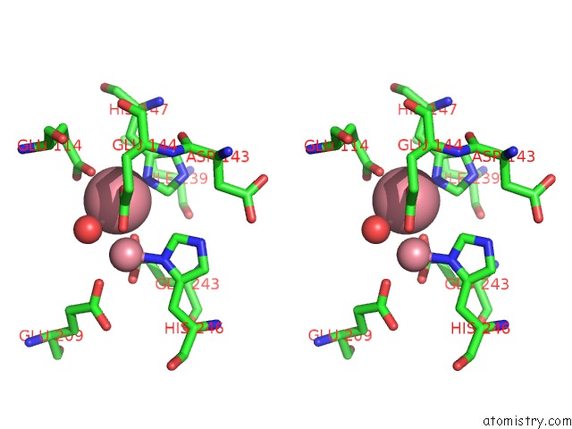
Cobalt binding site 1 out of 4 in 1xmh
Go back to
Cobalt binding site 1 out
of 4 in the Structure of Co(II) Reconstituted Methane Monooxygenase Hydroxylase From M. Capsulatus (Bath)

Mono view

Stereo pair view
Mono view
Stereo pair view
A full contact list of Cobalt with other atoms in the Co binding
site number 1 of Structure of Co(II) Reconstituted Methane Monooxygenase Hydroxylase From M. Capsulatus (Bath) within 5.0Å range:
|
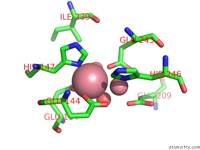
Cobalt binding site 2 out of 4 in 1xmh
Go back to
Cobalt binding site 2 out
of 4 in the Structure of Co(II) Reconstituted Methane Monooxygenase Hydroxylase From M. Capsulatus (Bath)

Mono view
Stereo pair view
Mono view
Stereo pair view
A full contact list of Cobalt with other atoms in the Co binding
site number 2 of Structure of Co(II) Reconstituted Methane Monooxygenase Hydroxylase From M. Capsulatus (Bath) within 5.0Å range:
|
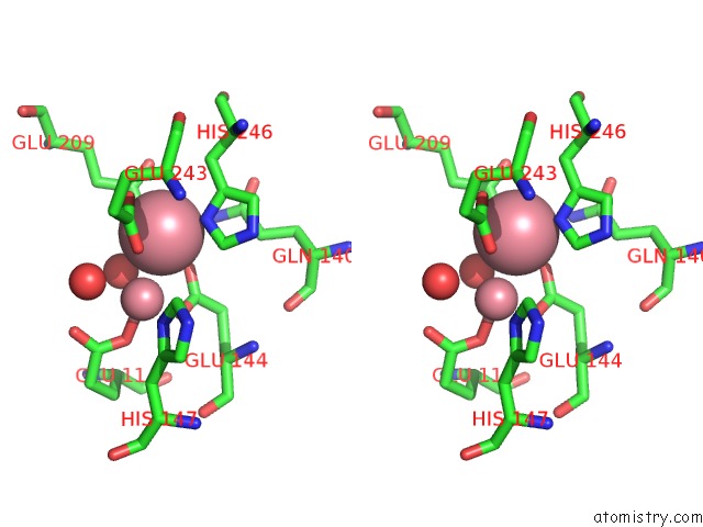
Cobalt binding site 3 out of 4 in 1xmh
Go back to
Cobalt binding site 3 out
of 4 in the Structure of Co(II) Reconstituted Methane Monooxygenase Hydroxylase From M. Capsulatus (Bath)
Mono view

Stereo pair view
Mono view
Stereo pair view
A full contact list of Cobalt with other atoms in the Co binding
site number 3 of Structure of Co(II) Reconstituted Methane Monooxygenase Hydroxylase From M. Capsulatus (Bath) within 5.0Å range:
|
Cobalt binding site 4 out of 4 in 1xmh
Go back to
Cobalt binding site 4 out
of 4 in the Structure of Co(II) Reconstituted Methane Monooxygenase Hydroxylase From M. Capsulatus (Bath)

Mono view
Stereo pair view
Mono view
Stereo pair view
A full contact list of Cobalt with other atoms in the Co binding
site number 4 of Structure of Co(II) Reconstituted Methane Monooxygenase Hydroxylase From M. Capsulatus (Bath) within 5.0Å range:
|
Reference:
M.H.Sazinsky,
M.Merkx,
E.Cadieux,
S.Tang,
S.J.Lippard.
Preparation and X-Ray Structures of Metal-Free, Dicobalt and Dimanganese Forms of Soluble Methane Monooxygenase Hydroxylase From Methylococcus Capsulatus (Bath) Biochemistry V. 43 16263 2004.
ISSN: ISSN 0006-2960
PubMed: 15610020
DOI: 10.1021/BI048140Z
Page generated: Tue Jul 30 14:46:33 2024
ISSN: ISSN 0006-2960
PubMed: 15610020
DOI: 10.1021/BI048140Z
Last articles
Zn in 9MJ5Zn in 9HNW
Zn in 9G0L
Zn in 9FNE
Zn in 9DZN
Zn in 9E0I
Zn in 9D32
Zn in 9DAK
Zn in 8ZXC
Zn in 8ZUF