Cobalt »
PDB 1vz0-1zft »
1xrs »
Cobalt in PDB 1xrs: Crystal Structure of Lysine 5,6-Aminomutase in Complex with Plp, Cobalamin, and 5'-Deoxyadenosine
Enzymatic activity of Crystal Structure of Lysine 5,6-Aminomutase in Complex with Plp, Cobalamin, and 5'-Deoxyadenosine
All present enzymatic activity of Crystal Structure of Lysine 5,6-Aminomutase in Complex with Plp, Cobalamin, and 5'-Deoxyadenosine:
5.4.3.3;
5.4.3.3;
Protein crystallography data
The structure of Crystal Structure of Lysine 5,6-Aminomutase in Complex with Plp, Cobalamin, and 5'-Deoxyadenosine, PDB code: 1xrs
was solved by
F.Berkovitch,
E.Behshad,
K.H.Tang,
E.A.Enns,
P.A.Frey,
C.L.Drennan,
with X-Ray Crystallography technique. A brief refinement statistics is given in the table below:
| Resolution Low / High (Å) | 49.85 / 2.80 |
| Space group | P 31 2 1 |
| Cell size a, b, c (Å), α, β, γ (°) | 99.700, 99.700, 168.800, 90.00, 90.00, 120.00 |
| R / Rfree (%) | 19.9 / 26.2 |
Cobalt Binding Sites:
The binding sites of Cobalt atom in the Crystal Structure of Lysine 5,6-Aminomutase in Complex with Plp, Cobalamin, and 5'-Deoxyadenosine
(pdb code 1xrs). This binding sites where shown within
5.0 Angstroms radius around Cobalt atom.
In total only one binding site of Cobalt was determined in the Crystal Structure of Lysine 5,6-Aminomutase in Complex with Plp, Cobalamin, and 5'-Deoxyadenosine, PDB code: 1xrs:
In total only one binding site of Cobalt was determined in the Crystal Structure of Lysine 5,6-Aminomutase in Complex with Plp, Cobalamin, and 5'-Deoxyadenosine, PDB code: 1xrs:
Cobalt binding site 1 out of 1 in 1xrs
Go back to
Cobalt binding site 1 out
of 1 in the Crystal Structure of Lysine 5,6-Aminomutase in Complex with Plp, Cobalamin, and 5'-Deoxyadenosine
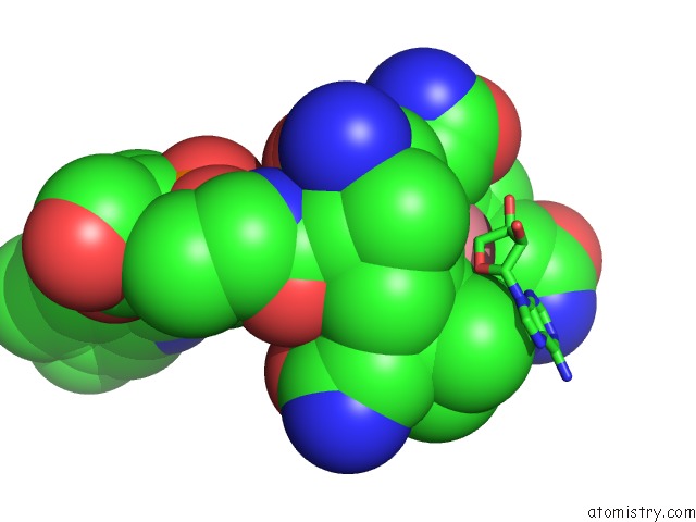
Mono view
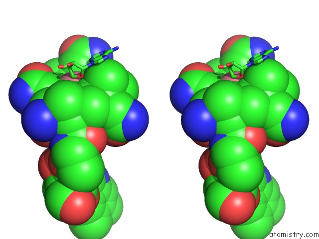
Stereo pair view
Mono view
Stereo pair view
A full contact list of Cobalt with other atoms in the Co binding
site number 1 of Crystal Structure of Lysine 5,6-Aminomutase in Complex with Plp, Cobalamin, and 5'-Deoxyadenosine within 5.0Å range:
|
Reference:
F.Berkovitch,
E.Behshad,
K.H.Tang,
E.A.Enns,
P.A.Frey,
C.L.Drennan.
A Locking Mechanism Preventing Radical Damage in the Absence of Substrate, As Revealed By the X-Ray Structure of Lysine 5,6-Aminomutase. Proc.Natl.Acad.Sci.Usa V. 101 15870 2004.
ISSN: ISSN 0027-8424
PubMed: 15514022
DOI: 10.1073/PNAS.0407074101
Page generated: Tue Jul 30 14:48:12 2024
ISSN: ISSN 0027-8424
PubMed: 15514022
DOI: 10.1073/PNAS.0407074101
Last articles
Zn in 9J0NZn in 9J0O
Zn in 9J0P
Zn in 9FJX
Zn in 9EKB
Zn in 9C0F
Zn in 9CAH
Zn in 9CH0
Zn in 9CH3
Zn in 9CH1