Cobalt »
PDB 2djl-2g6p »
2e6a »
Cobalt in PDB 2e6a: Crystal Structure of Trypanosoma Cruzi Dihydroorotate Dehydrogenase in Complex with Orotate
Enzymatic activity of Crystal Structure of Trypanosoma Cruzi Dihydroorotate Dehydrogenase in Complex with Orotate
All present enzymatic activity of Crystal Structure of Trypanosoma Cruzi Dihydroorotate Dehydrogenase in Complex with Orotate:
1.3.3.1;
1.3.3.1;
Protein crystallography data
The structure of Crystal Structure of Trypanosoma Cruzi Dihydroorotate Dehydrogenase in Complex with Orotate, PDB code: 2e6a
was solved by
D.K.Inaoka,
H.Shimizu,
K.Sakamoto,
T.Shiba,
G.Kurisu,
T.Nara,
T.Aoki,
S.Harada,
K.Kita,
with X-Ray Crystallography technique. A brief refinement statistics is given in the table below:
| Resolution Low / High (Å) | 49.51 / 1.64 |
| Space group | P 21 21 21 |
| Cell size a, b, c (Å), α, β, γ (°) | 68.248, 71.881, 123.574, 90.00, 90.00, 90.00 |
| R / Rfree (%) | 16.2 / 19.2 |
Cobalt Binding Sites:
The binding sites of Cobalt atom in the Crystal Structure of Trypanosoma Cruzi Dihydroorotate Dehydrogenase in Complex with Orotate
(pdb code 2e6a). This binding sites where shown within
5.0 Angstroms radius around Cobalt atom.
In total only one binding site of Cobalt was determined in the Crystal Structure of Trypanosoma Cruzi Dihydroorotate Dehydrogenase in Complex with Orotate, PDB code: 2e6a:
In total only one binding site of Cobalt was determined in the Crystal Structure of Trypanosoma Cruzi Dihydroorotate Dehydrogenase in Complex with Orotate, PDB code: 2e6a:
Cobalt binding site 1 out of 1 in 2e6a
Go back to
Cobalt binding site 1 out
of 1 in the Crystal Structure of Trypanosoma Cruzi Dihydroorotate Dehydrogenase in Complex with Orotate
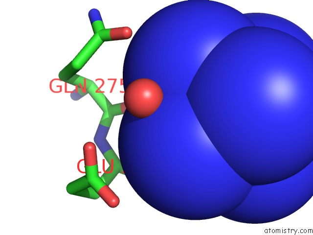
Mono view
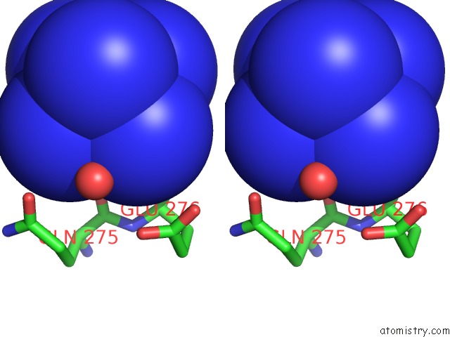
Stereo pair view
Mono view
Stereo pair view
A full contact list of Cobalt with other atoms in the Co binding
site number 1 of Crystal Structure of Trypanosoma Cruzi Dihydroorotate Dehydrogenase in Complex with Orotate within 5.0Å range:
|
Reference:
D.K.Inaoka,
H.Shimizu,
K.Sakamoto,
T.Shiba,
G.Kurisu,
T.Nara,
T.Aoki,
S.Harada,
K.Kita.
Crystal Structure of Trypanosoma Cruzi Dihydroorotate Dehydrogenase in Complex with Orotate To Be Published.
Page generated: Sun Jul 13 18:08:48 2025
Last articles
Cu in 4YL4Cu in 4XQZ
Cu in 4YEA
Cu in 4XYD
Cu in 4X4K
Cu in 4XSN
Cu in 4WKX
Cu in 4X27
Cu in 4WZ9
Cu in 4W9Z