Cobalt »
PDB 2djl-2g6p »
2f7v »
Cobalt in PDB 2f7v: Structure of Acetylcitrulline Deacetylase Complexed with One Co
Enzymatic activity of Structure of Acetylcitrulline Deacetylase Complexed with One Co
All present enzymatic activity of Structure of Acetylcitrulline Deacetylase Complexed with One Co:
3.5.1.16;
3.5.1.16;
Protein crystallography data
The structure of Structure of Acetylcitrulline Deacetylase Complexed with One Co, PDB code: 2f7v
was solved by
D.Shi,
X.Yu,
L.Roth,
N.M.Allewell,
M.Tuchman,
with X-Ray Crystallography technique. A brief refinement statistics is given in the table below:
| Resolution Low / High (Å) | 37.33 / 1.75 |
| Space group | C 1 2 1 |
| Cell size a, b, c (Å), α, β, γ (°) | 94.068, 95.429, 43.670, 90.00, 93.77, 90.00 |
| R / Rfree (%) | 19.7 / 22.5 |
Cobalt Binding Sites:
The binding sites of Cobalt atom in the Structure of Acetylcitrulline Deacetylase Complexed with One Co
(pdb code 2f7v). This binding sites where shown within
5.0 Angstroms radius around Cobalt atom.
In total only one binding site of Cobalt was determined in the Structure of Acetylcitrulline Deacetylase Complexed with One Co, PDB code: 2f7v:
In total only one binding site of Cobalt was determined in the Structure of Acetylcitrulline Deacetylase Complexed with One Co, PDB code: 2f7v:
Cobalt binding site 1 out of 1 in 2f7v
Go back to
Cobalt binding site 1 out
of 1 in the Structure of Acetylcitrulline Deacetylase Complexed with One Co
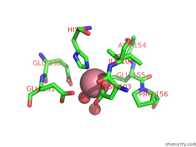
Mono view
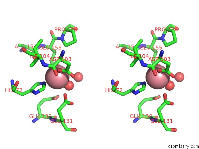
Stereo pair view
Mono view
Stereo pair view
A full contact list of Cobalt with other atoms in the Co binding
site number 1 of Structure of Acetylcitrulline Deacetylase Complexed with One Co within 5.0Å range:
|
Reference:
D.Shi,
X.Yu,
L.Roth,
M.Tuchman,
N.M.Allewell.
Structure of A Novel N-Acetyl-L-Citrulline Deacetylase From Xanthomonas Campestris Biophys.Chem. V. 126 86 2007.
ISSN: ISSN 0301-4622
PubMed: 16750290
DOI: 10.1016/J.BPC.2006.05.013
Page generated: Sun Jul 13 18:13:34 2025
ISSN: ISSN 0301-4622
PubMed: 16750290
DOI: 10.1016/J.BPC.2006.05.013
Last articles
Cu in 3MLJCu in 3MLK
Cu in 3MLL
Cu in 3MIG
Cu in 3MIH
Cu in 3MIF
Cu in 3MIE
Cu in 3MID
Cu in 3MIC
Cu in 3MIB