Cobalt »
PDB 2djl-2g6p »
2fdf »
Cobalt in PDB 2fdf: Crystal Structure of Alkb in Complex with Co(II), 2-Oxoglutarate, and Methylated Trinucleotide T-Mea-T
Protein crystallography data
The structure of Crystal Structure of Alkb in Complex with Co(II), 2-Oxoglutarate, and Methylated Trinucleotide T-Mea-T, PDB code: 2fdf
was solved by
B.Yu,
J.Benach,
W.C.Edstrom,
B.R.Gibney,
J.F.Hunt,
Northeast Structuralgenomics Consortium (Nesg),
with X-Ray Crystallography technique. A brief refinement statistics is given in the table below:
| Resolution Low / High (Å) | 19.47 / 2.10 |
| Space group | P 43 |
| Cell size a, b, c (Å), α, β, γ (°) | 41.294, 41.294, 116.797, 90.00, 90.00, 90.00 |
| R / Rfree (%) | 19.3 / 23.7 |
Cobalt Binding Sites:
The binding sites of Cobalt atom in the Crystal Structure of Alkb in Complex with Co(II), 2-Oxoglutarate, and Methylated Trinucleotide T-Mea-T
(pdb code 2fdf). This binding sites where shown within
5.0 Angstroms radius around Cobalt atom.
In total only one binding site of Cobalt was determined in the Crystal Structure of Alkb in Complex with Co(II), 2-Oxoglutarate, and Methylated Trinucleotide T-Mea-T, PDB code: 2fdf:
In total only one binding site of Cobalt was determined in the Crystal Structure of Alkb in Complex with Co(II), 2-Oxoglutarate, and Methylated Trinucleotide T-Mea-T, PDB code: 2fdf:
Cobalt binding site 1 out of 1 in 2fdf
Go back to
Cobalt binding site 1 out
of 1 in the Crystal Structure of Alkb in Complex with Co(II), 2-Oxoglutarate, and Methylated Trinucleotide T-Mea-T
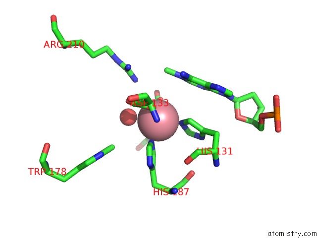
Mono view
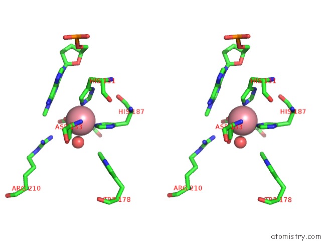
Stereo pair view
Mono view
Stereo pair view
A full contact list of Cobalt with other atoms in the Co binding
site number 1 of Crystal Structure of Alkb in Complex with Co(II), 2-Oxoglutarate, and Methylated Trinucleotide T-Mea-T within 5.0Å range:
|
Reference:
B.Yu,
W.C.Edstrom,
J.Benach,
Y.Hamuro,
P.C.Weber,
B.R.Gibney,
J.F.Hunt.
Crystal Structures of Catalytic Complexes of the Oxidative Dna/Rna Repair Enzyme Alkb. Nature V. 439 879 2006.
ISSN: ISSN 0028-0836
PubMed: 16482161
DOI: 10.1038/NATURE04561
Page generated: Sun Jul 13 18:13:41 2025
ISSN: ISSN 0028-0836
PubMed: 16482161
DOI: 10.1038/NATURE04561
Last articles
Cu in 3T6VCu in 3SBR
Cu in 3SBP
Cu in 3T6Q
Cu in 3T5W
Cu in 3T56
Cu in 3T53
Cu in 3T51
Cu in 3T0U
Cu in 3SQR