Cobalt »
PDB 2g9c-2nq7 »
2gve »
Cobalt in PDB 2gve: Time-of-Flight Neutron Diffraction Structure of D-Xylose Isomerase
Enzymatic activity of Time-of-Flight Neutron Diffraction Structure of D-Xylose Isomerase
All present enzymatic activity of Time-of-Flight Neutron Diffraction Structure of D-Xylose Isomerase:
5.3.1.5;
5.3.1.5;
Cobalt Binding Sites:
The binding sites of Cobalt atom in the Time-of-Flight Neutron Diffraction Structure of D-Xylose Isomerase
(pdb code 2gve). This binding sites where shown within
5.0 Angstroms radius around Cobalt atom.
In total 2 binding sites of Cobalt where determined in the Time-of-Flight Neutron Diffraction Structure of D-Xylose Isomerase, PDB code: 2gve:
Jump to Cobalt binding site number: 1; 2;
In total 2 binding sites of Cobalt where determined in the Time-of-Flight Neutron Diffraction Structure of D-Xylose Isomerase, PDB code: 2gve:
Jump to Cobalt binding site number: 1; 2;
Cobalt binding site 1 out of 2 in 2gve
Go back to
Cobalt binding site 1 out
of 2 in the Time-of-Flight Neutron Diffraction Structure of D-Xylose Isomerase
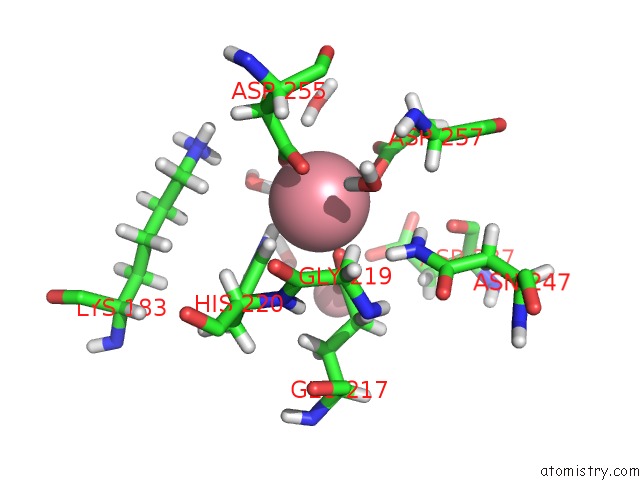
Mono view
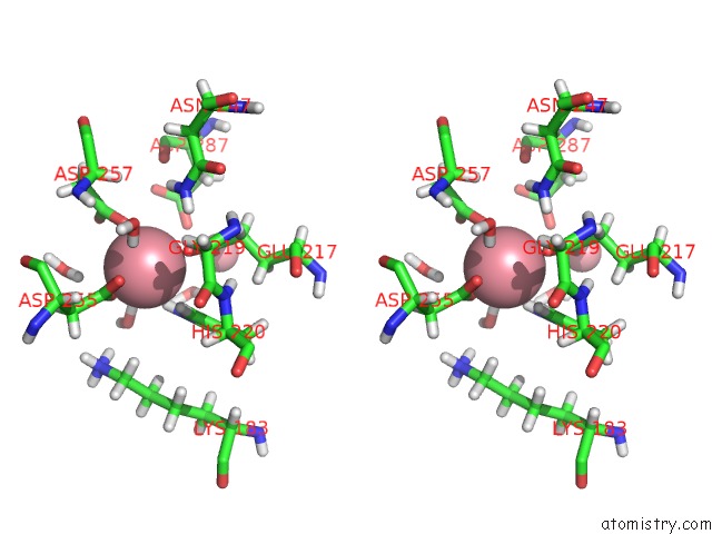
Stereo pair view
Mono view
Stereo pair view
A full contact list of Cobalt with other atoms in the Co binding
site number 1 of Time-of-Flight Neutron Diffraction Structure of D-Xylose Isomerase within 5.0Å range:
|
Cobalt binding site 2 out of 2 in 2gve
Go back to
Cobalt binding site 2 out
of 2 in the Time-of-Flight Neutron Diffraction Structure of D-Xylose Isomerase
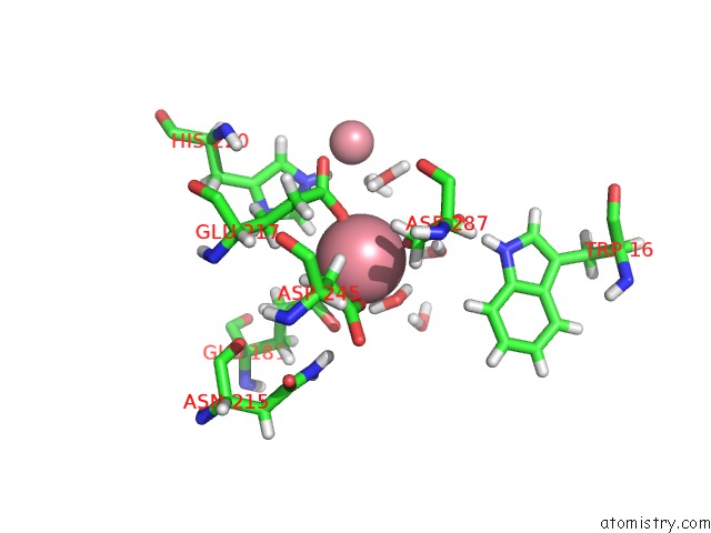
Mono view
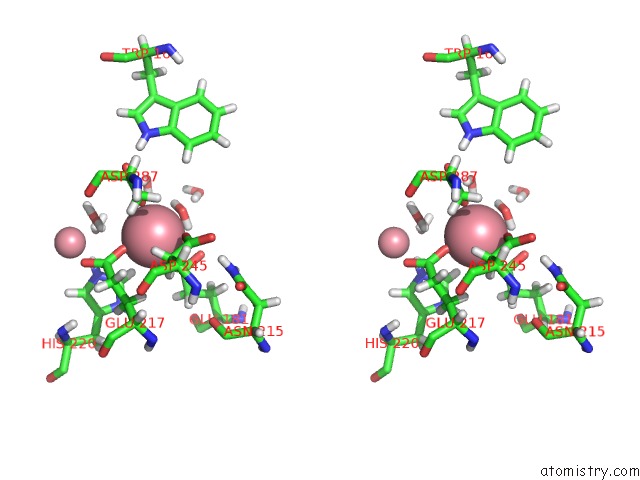
Stereo pair view
Mono view
Stereo pair view
A full contact list of Cobalt with other atoms in the Co binding
site number 2 of Time-of-Flight Neutron Diffraction Structure of D-Xylose Isomerase within 5.0Å range:
|
Reference:
A.K.Katz,
X.Li,
H.L.Carrell,
B.L.Hanson,
P.Langan,
L.Coates,
B.P.Schoenborn,
J.P.Glusker,
G.J.Bunick.
Locating Active-Site Hydrogen Atoms in D-Xylose Isomerase: Time-of-Flight Neutron Diffraction. Proc.Natl.Acad.Sci.Usa V. 103 8342 2006.
ISSN: ISSN 0027-8424
PubMed: 16707576
DOI: 10.1073/PNAS.0602598103
Page generated: Tue Jul 30 15:14:42 2024
ISSN: ISSN 0027-8424
PubMed: 16707576
DOI: 10.1073/PNAS.0602598103
Last articles
Zn in 9J0NZn in 9J0O
Zn in 9J0P
Zn in 9FJX
Zn in 9EKB
Zn in 9C0F
Zn in 9CAH
Zn in 9CH0
Zn in 9CH3
Zn in 9CH1