Cobalt »
PDB 2g9c-2nq7 »
2mat »
Cobalt in PDB 2mat: E.Coli Methionine Aminopeptidase at 1.9 Angstrom Resolution
Enzymatic activity of E.Coli Methionine Aminopeptidase at 1.9 Angstrom Resolution
All present enzymatic activity of E.Coli Methionine Aminopeptidase at 1.9 Angstrom Resolution:
3.4.11.18;
3.4.11.18;
Protein crystallography data
The structure of E.Coli Methionine Aminopeptidase at 1.9 Angstrom Resolution, PDB code: 2mat
was solved by
W.T.Lowther,
A.M.Orville,
D.T.Madden,
S.Lim,
D.H.Rich,
B.W.Matthews,
with X-Ray Crystallography technique. A brief refinement statistics is given in the table below:
| Resolution Low / High (Å) | 35.50 / 1.90 |
| Space group | P 1 21 1 |
| Cell size a, b, c (Å), α, β, γ (°) | 39.299, 67.683, 48.863, 90.00, 111.24, 90.00 |
| R / Rfree (%) | n/a / n/a |
Other elements in 2mat:
The structure of E.Coli Methionine Aminopeptidase at 1.9 Angstrom Resolution also contains other interesting chemical elements:
| Sodium | (Na) | 1 atom |
Cobalt Binding Sites:
The binding sites of Cobalt atom in the E.Coli Methionine Aminopeptidase at 1.9 Angstrom Resolution
(pdb code 2mat). This binding sites where shown within
5.0 Angstroms radius around Cobalt atom.
In total 3 binding sites of Cobalt where determined in the E.Coli Methionine Aminopeptidase at 1.9 Angstrom Resolution, PDB code: 2mat:
Jump to Cobalt binding site number: 1; 2; 3;
In total 3 binding sites of Cobalt where determined in the E.Coli Methionine Aminopeptidase at 1.9 Angstrom Resolution, PDB code: 2mat:
Jump to Cobalt binding site number: 1; 2; 3;
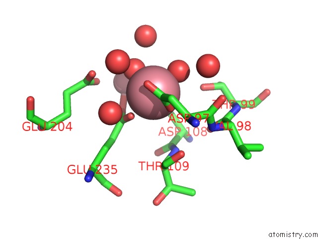
Cobalt binding site 1 out of 3 in 2mat
Go back to
Cobalt binding site 1 out
of 3 in the E.Coli Methionine Aminopeptidase at 1.9 Angstrom Resolution

Mono view

Stereo pair view
Mono view
Stereo pair view
A full contact list of Cobalt with other atoms in the Co binding
site number 1 of E.Coli Methionine Aminopeptidase at 1.9 Angstrom Resolution within 5.0Å range:
|
Cobalt binding site 2 out of 3 in 2mat
Go back to
Cobalt binding site 2 out
of 3 in the E.Coli Methionine Aminopeptidase at 1.9 Angstrom Resolution
Mono view

Stereo pair view
Mono view
Stereo pair view
A full contact list of Cobalt with other atoms in the Co binding
site number 2 of E.Coli Methionine Aminopeptidase at 1.9 Angstrom Resolution within 5.0Å range:
|
Cobalt binding site 3 out of 3 in 2mat
Go back to
Cobalt binding site 3 out
of 3 in the E.Coli Methionine Aminopeptidase at 1.9 Angstrom Resolution

Mono view

Stereo pair view
Mono view
Stereo pair view
A full contact list of Cobalt with other atoms in the Co binding
site number 3 of E.Coli Methionine Aminopeptidase at 1.9 Angstrom Resolution within 5.0Å range:
|
Reference:
W.T.Lowther,
A.M.Orville,
D.T.Madden,
S.Lim,
D.H.Rich,
B.W.Matthews.
Escherichia Coli Methionine Aminopeptidase: Implications of Crystallographic Analyses of the Native, Mutant, and Inhibited Enzymes For the Mechanism of Catalysis. Biochemistry V. 38 7678 1999.
ISSN: ISSN 0006-2960
PubMed: 10387007
DOI: 10.1021/BI990684R
Page generated: Tue Jul 30 15:17:58 2024
ISSN: ISSN 0006-2960
PubMed: 10387007
DOI: 10.1021/BI990684R
Last articles
Zn in 9J0NZn in 9J0O
Zn in 9J0P
Zn in 9FJX
Zn in 9EKB
Zn in 9C0F
Zn in 9CAH
Zn in 9CH0
Zn in 9CH3
Zn in 9CH1