Cobalt »
PDB 2xwq-3bbi »
2zu9 »
Cobalt in PDB 2zu9: Crystal Structure of Mannosyl-3-Phosphoglycerate Synthase From Pyrococcus Horikoshii
Enzymatic activity of Crystal Structure of Mannosyl-3-Phosphoglycerate Synthase From Pyrococcus Horikoshii
All present enzymatic activity of Crystal Structure of Mannosyl-3-Phosphoglycerate Synthase From Pyrococcus Horikoshii:
2.4.1.217;
2.4.1.217;
Protein crystallography data
The structure of Crystal Structure of Mannosyl-3-Phosphoglycerate Synthase From Pyrococcus Horikoshii, PDB code: 2zu9
was solved by
T.Kawamura,
N.Watanabe,
I.Tanaka,
with X-Ray Crystallography technique. A brief refinement statistics is given in the table below:
| Resolution Low / High (Å) | 42.91 / 2.00 |
| Space group | P 1 21 1 |
| Cell size a, b, c (Å), α, β, γ (°) | 54.655, 84.867, 83.565, 90.00, 102.00, 90.00 |
| R / Rfree (%) | 22.4 / 25.6 |
Cobalt Binding Sites:
The binding sites of Cobalt atom in the Crystal Structure of Mannosyl-3-Phosphoglycerate Synthase From Pyrococcus Horikoshii
(pdb code 2zu9). This binding sites where shown within
5.0 Angstroms radius around Cobalt atom.
In total 2 binding sites of Cobalt where determined in the Crystal Structure of Mannosyl-3-Phosphoglycerate Synthase From Pyrococcus Horikoshii, PDB code: 2zu9:
Jump to Cobalt binding site number: 1; 2;
In total 2 binding sites of Cobalt where determined in the Crystal Structure of Mannosyl-3-Phosphoglycerate Synthase From Pyrococcus Horikoshii, PDB code: 2zu9:
Jump to Cobalt binding site number: 1; 2;
Cobalt binding site 1 out of 2 in 2zu9
Go back to
Cobalt binding site 1 out
of 2 in the Crystal Structure of Mannosyl-3-Phosphoglycerate Synthase From Pyrococcus Horikoshii
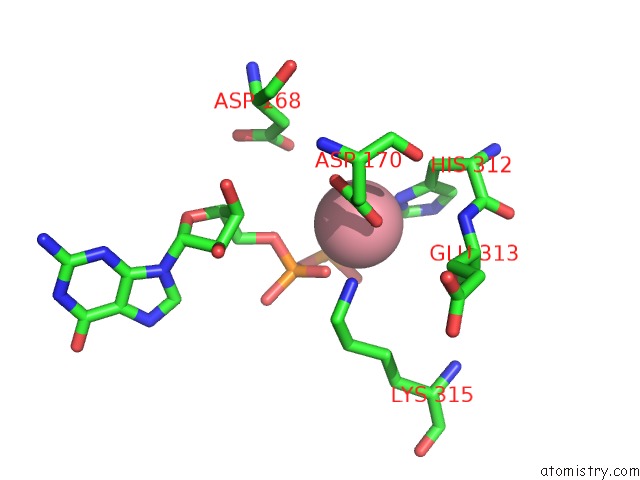
Mono view
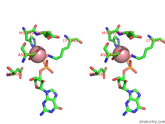
Stereo pair view
Mono view
Stereo pair view
A full contact list of Cobalt with other atoms in the Co binding
site number 1 of Crystal Structure of Mannosyl-3-Phosphoglycerate Synthase From Pyrococcus Horikoshii within 5.0Å range:
|
Cobalt binding site 2 out of 2 in 2zu9
Go back to
Cobalt binding site 2 out
of 2 in the Crystal Structure of Mannosyl-3-Phosphoglycerate Synthase From Pyrococcus Horikoshii
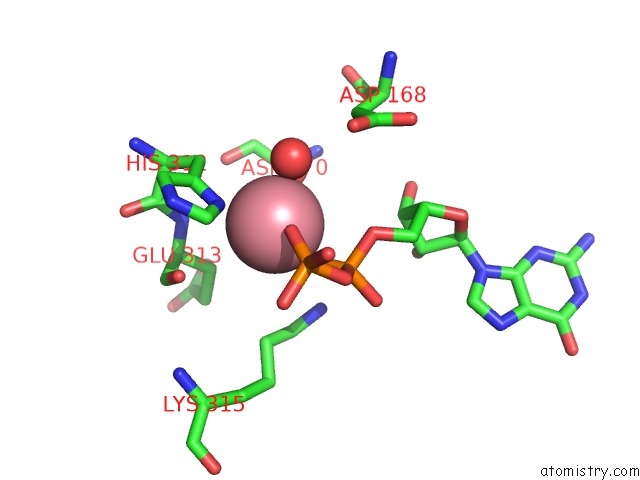
Mono view

Stereo pair view
Mono view
Stereo pair view
A full contact list of Cobalt with other atoms in the Co binding
site number 2 of Crystal Structure of Mannosyl-3-Phosphoglycerate Synthase From Pyrococcus Horikoshii within 5.0Å range:
|
Reference:
T.Kawamura,
N.Watanabe,
I.Tanaka.
Crystal Structure of Mannosyl-3-Phosphoglycerate Synthase From Pyrococcus Horikoshii To Be Published.
Page generated: Sun Jul 13 18:40:51 2025
Last articles
Cu in 5I26Cu in 5I6L
Cu in 5I38
Cu in 5I6K
Cu in 5HZT
Cu in 5FJE
Cu in 5I0Y
Cu in 5I0X
Cu in 5I0W
Cu in 5I0V