Cobalt »
PDB 2xwq-3bbi »
3a9i »
Cobalt in PDB 3a9i: Crystal Structure of Homocitrate Synthase From Thermus Thermophilus Complexed with Lys
Enzymatic activity of Crystal Structure of Homocitrate Synthase From Thermus Thermophilus Complexed with Lys
All present enzymatic activity of Crystal Structure of Homocitrate Synthase From Thermus Thermophilus Complexed with Lys:
2.3.3.14;
2.3.3.14;
Protein crystallography data
The structure of Crystal Structure of Homocitrate Synthase From Thermus Thermophilus Complexed with Lys, PDB code: 3a9i
was solved by
T.Okada,
T.Tomita,
T.Kuzuyama,
M.Nishiyama,
with X-Ray Crystallography technique. A brief refinement statistics is given in the table below:
| Resolution Low / High (Å) | 35.60 / 1.80 |
| Space group | C 2 2 21 |
| Cell size a, b, c (Å), α, β, γ (°) | 80.281, 96.025, 77.082, 90.00, 90.00, 90.00 |
| R / Rfree (%) | 17.1 / 21.3 |
Cobalt Binding Sites:
The binding sites of Cobalt atom in the Crystal Structure of Homocitrate Synthase From Thermus Thermophilus Complexed with Lys
(pdb code 3a9i). This binding sites where shown within
5.0 Angstroms radius around Cobalt atom.
In total only one binding site of Cobalt was determined in the Crystal Structure of Homocitrate Synthase From Thermus Thermophilus Complexed with Lys, PDB code: 3a9i:
In total only one binding site of Cobalt was determined in the Crystal Structure of Homocitrate Synthase From Thermus Thermophilus Complexed with Lys, PDB code: 3a9i:
Cobalt binding site 1 out of 1 in 3a9i
Go back to
Cobalt binding site 1 out
of 1 in the Crystal Structure of Homocitrate Synthase From Thermus Thermophilus Complexed with Lys
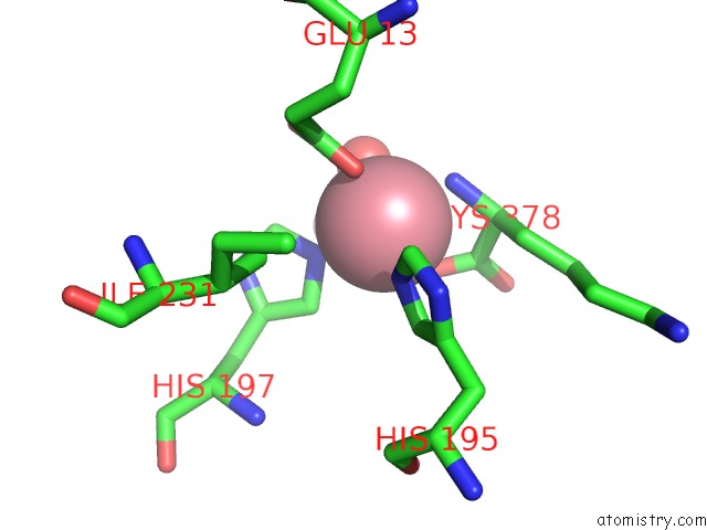
Mono view
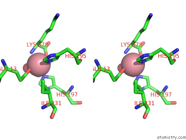
Stereo pair view
Mono view
Stereo pair view
A full contact list of Cobalt with other atoms in the Co binding
site number 1 of Crystal Structure of Homocitrate Synthase From Thermus Thermophilus Complexed with Lys within 5.0Å range:
|
Reference:
T.Okada,
T.Tomita,
A.P.Wulandari,
T.Kuzuyama,
M.Nishiyama.
Mechanism of Substrate Recognition and Insight Into Feedback Inhibition of Homocitrate Synthase From Thermus Thermophilus J.Biol.Chem. V. 285 4195 2010.
ISSN: ISSN 0021-9258
PubMed: 19996101
DOI: 10.1074/JBC.M109.086330
Page generated: Sun Jul 13 18:42:21 2025
ISSN: ISSN 0021-9258
PubMed: 19996101
DOI: 10.1074/JBC.M109.086330
Last articles
Cu in 4Z0ZCu in 4YSU
Cu in 4YZW
Cu in 4YVU
Cu in 4YVN
Cu in 4YSR
Cu in 4YSS
Cu in 4YST
Cu in 4YSQ
Cu in 4YSD