Cobalt »
PDB 3bbk-3ger »
3bfq »
Cobalt in PDB 3bfq: Crystal Structure of Truncated Fimg (Fimgt) in Complex with the Donor Strand Peptide of Fimf (Dsf)
Protein crystallography data
The structure of Crystal Structure of Truncated Fimg (Fimgt) in Complex with the Donor Strand Peptide of Fimf (Dsf), PDB code: 3bfq
was solved by
O.Eidam,
G.Capitani,
M.G.Grutter,
with X-Ray Crystallography technique. A brief refinement statistics is given in the table below:
| Resolution Low / High (Å) | 15.00 / 1.34 |
| Space group | P 1 21 1 |
| Cell size a, b, c (Å), α, β, γ (°) | 48.050, 24.500, 54.150, 90.00, 113.77, 90.00 |
| R / Rfree (%) | 13.2 / 17.9 |
Cobalt Binding Sites:
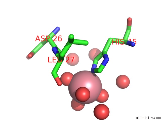
The binding sites of Cobalt atom in the Crystal Structure of Truncated Fimg (Fimgt) in Complex with the Donor Strand Peptide of Fimf (Dsf)
(pdb code 3bfq). This binding sites where shown within
5.0 Angstroms radius around Cobalt atom.
In total only one binding site of Cobalt was determined in the Crystal Structure of Truncated Fimg (Fimgt) in Complex with the Donor Strand Peptide of Fimf (Dsf), PDB code: 3bfq:
In total only one binding site of Cobalt was determined in the Crystal Structure of Truncated Fimg (Fimgt) in Complex with the Donor Strand Peptide of Fimf (Dsf), PDB code: 3bfq:
Cobalt binding site 1 out of 1 in 3bfq
Go back to
Cobalt binding site 1 out
of 1 in the Crystal Structure of Truncated Fimg (Fimgt) in Complex with the Donor Strand Peptide of Fimf (Dsf)
Mono view
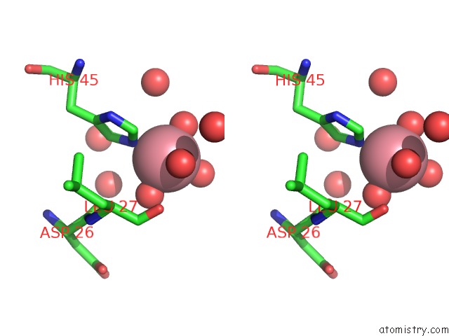
Stereo pair view
Mono view
Stereo pair view
A full contact list of Cobalt with other atoms in the Co binding
site number 1 of Crystal Structure of Truncated Fimg (Fimgt) in Complex with the Donor Strand Peptide of Fimf (Dsf) within 5.0Å range:
|
Reference:
C.Puorger,
O.Eidam,
G.Capitani,
D.Erilov,
M.G.Grutter,
R.Glockshuber.
Infinite Kinetic Stability Against Dissociation of Supramolecular Protein Complexes Through Donor Strand Complementation Structure V. 16 631 2008.
ISSN: ISSN 0969-2126
PubMed: 18400183
DOI: 10.1016/J.STR.2008.01.013
Page generated: Sun Jul 13 18:45:22 2025
ISSN: ISSN 0969-2126
PubMed: 18400183
DOI: 10.1016/J.STR.2008.01.013
Last articles
Cu in 3ZBMCu in 3X42
Cu in 3X41
Cu in 3X40
Cu in 3X3Z
Cu in 3X3X
Cu in 3X3Y
Cu in 3X1G
Cu in 3X1F
Cu in 3X1N