Cobalt »
PDB 3bbk-3ger »
3g4m »
Cobalt in PDB 3g4m: Crystal Structure of Guanine Riboswitch Bound to 2- Aminopurine
Protein crystallography data
The structure of Crystal Structure of Guanine Riboswitch Bound to 2- Aminopurine, PDB code: 3g4m
was solved by
S.D.Gilbert,
R.T.Batey,
with X-Ray Crystallography technique. A brief refinement statistics is given in the table below:
| Resolution Low / High (Å) | 19.92 / 2.40 |
| Space group | C 1 2 1 |
| Cell size a, b, c (Å), α, β, γ (°) | 127.430, 35.160, 42.240, 90.00, 91.18, 90.00 |
| R / Rfree (%) | 22.9 / 27.8 |
Cobalt Binding Sites:
Pages:
>>> Page 1 <<< Page 2, Binding sites: 11 - 11;Binding sites:
The binding sites of Cobalt atom in the Crystal Structure of Guanine Riboswitch Bound to 2- Aminopurine (pdb code 3g4m). This binding sites where shown within 5.0 Angstroms radius around Cobalt atom.In total 11 binding sites of Cobalt where determined in the Crystal Structure of Guanine Riboswitch Bound to 2- Aminopurine, PDB code: 3g4m:
Jump to Cobalt binding site number: 1; 2; 3; 4; 5; 6; 7; 8; 9; 10;
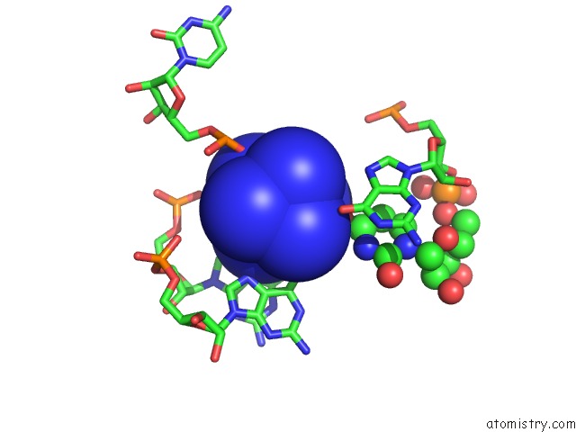
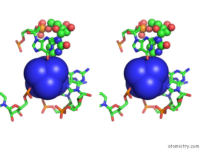
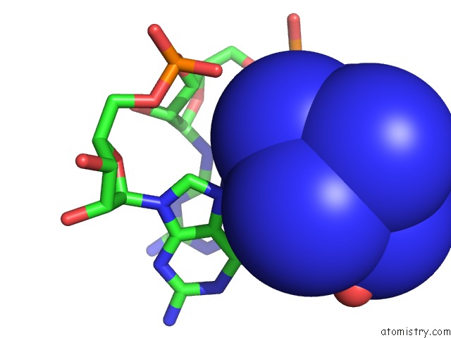
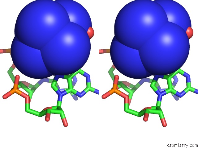
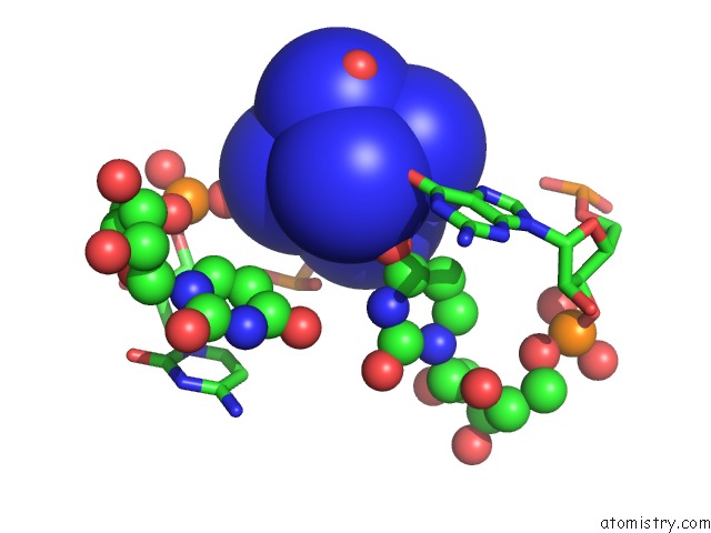
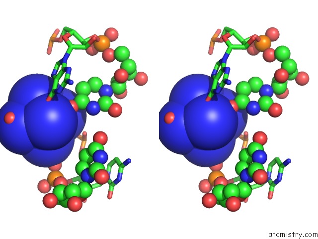
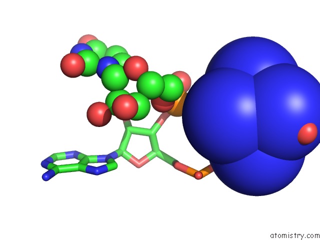
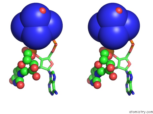
Cobalt binding site 1 out of 11 in 3g4m
Go back to
Cobalt binding site 1 out
of 11 in the Crystal Structure of Guanine Riboswitch Bound to 2- Aminopurine
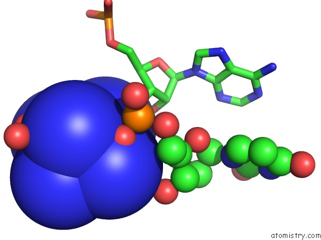
Mono view
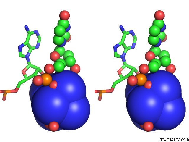
Stereo pair view
Mono view
Stereo pair view
A full contact list of Cobalt with other atoms in the Co binding
site number 1 of Crystal Structure of Guanine Riboswitch Bound to 2- Aminopurine within 5.0Å range:
|
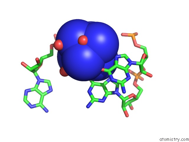
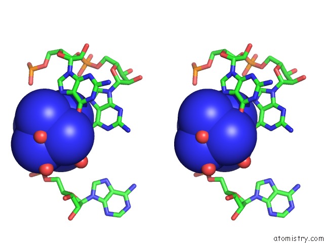
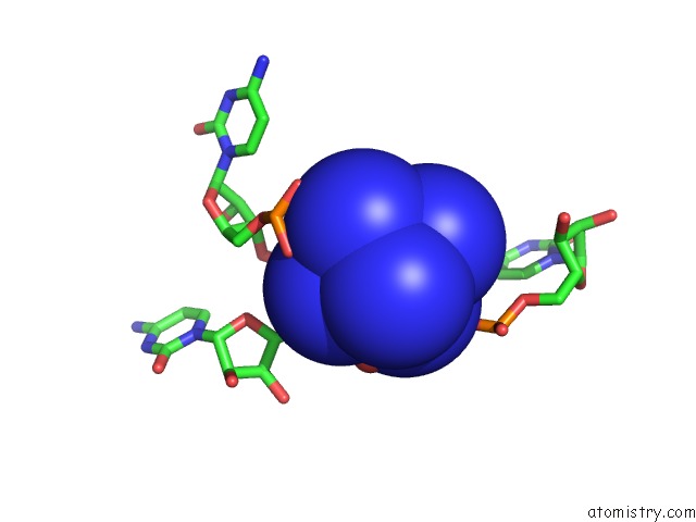
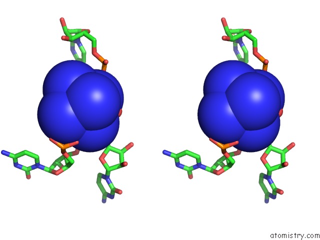
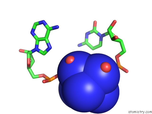
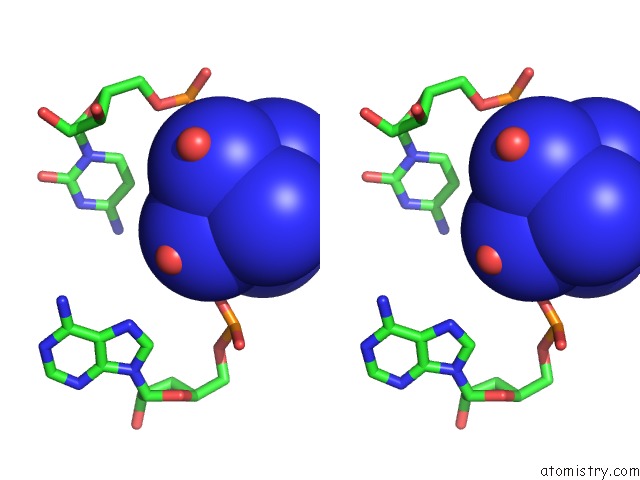
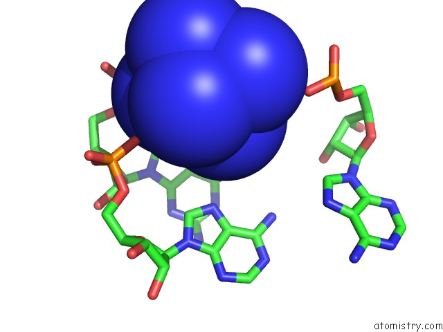
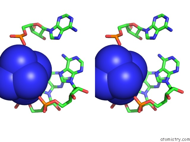
Cobalt binding site 2 out of 11 in 3g4m
Go back to
Cobalt binding site 2 out
of 11 in the Crystal Structure of Guanine Riboswitch Bound to 2- Aminopurine
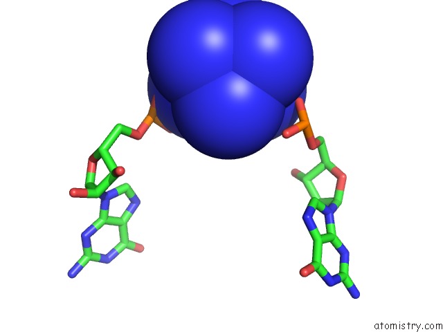
Mono view
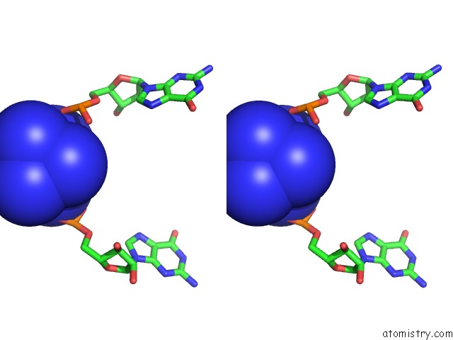
Stereo pair view
Mono view
Stereo pair view
A full contact list of Cobalt with other atoms in the Co binding
site number 2 of Crystal Structure of Guanine Riboswitch Bound to 2- Aminopurine within 5.0Å range:
|
Cobalt binding site 3 out of 11 in 3g4m
Go back to
Cobalt binding site 3 out
of 11 in the Crystal Structure of Guanine Riboswitch Bound to 2- Aminopurine
Mono view
Stereo pair view
Mono view
Stereo pair view
A full contact list of Cobalt with other atoms in the Co binding
site number 3 of Crystal Structure of Guanine Riboswitch Bound to 2- Aminopurine within 5.0Å range:
|
Cobalt binding site 4 out of 11 in 3g4m
Go back to
Cobalt binding site 4 out
of 11 in the Crystal Structure of Guanine Riboswitch Bound to 2- Aminopurine
Mono view
Stereo pair view
Mono view
Stereo pair view
A full contact list of Cobalt with other atoms in the Co binding
site number 4 of Crystal Structure of Guanine Riboswitch Bound to 2- Aminopurine within 5.0Å range:
|
Cobalt binding site 5 out of 11 in 3g4m
Go back to
Cobalt binding site 5 out
of 11 in the Crystal Structure of Guanine Riboswitch Bound to 2- Aminopurine
Mono view
Stereo pair view
Mono view
Stereo pair view
A full contact list of Cobalt with other atoms in the Co binding
site number 5 of Crystal Structure of Guanine Riboswitch Bound to 2- Aminopurine within 5.0Å range:
|
Cobalt binding site 6 out of 11 in 3g4m
Go back to
Cobalt binding site 6 out
of 11 in the Crystal Structure of Guanine Riboswitch Bound to 2- Aminopurine
Mono view
Stereo pair view
Mono view
Stereo pair view
A full contact list of Cobalt with other atoms in the Co binding
site number 6 of Crystal Structure of Guanine Riboswitch Bound to 2- Aminopurine within 5.0Å range:
|
Cobalt binding site 7 out of 11 in 3g4m
Go back to
Cobalt binding site 7 out
of 11 in the Crystal Structure of Guanine Riboswitch Bound to 2- Aminopurine
Mono view
Stereo pair view
Mono view
Stereo pair view
A full contact list of Cobalt with other atoms in the Co binding
site number 7 of Crystal Structure of Guanine Riboswitch Bound to 2- Aminopurine within 5.0Å range:
|
Cobalt binding site 8 out of 11 in 3g4m
Go back to
Cobalt binding site 8 out
of 11 in the Crystal Structure of Guanine Riboswitch Bound to 2- Aminopurine
Mono view
Stereo pair view
Mono view
Stereo pair view
A full contact list of Cobalt with other atoms in the Co binding
site number 8 of Crystal Structure of Guanine Riboswitch Bound to 2- Aminopurine within 5.0Å range:
|
Cobalt binding site 9 out of 11 in 3g4m
Go back to
Cobalt binding site 9 out
of 11 in the Crystal Structure of Guanine Riboswitch Bound to 2- Aminopurine
Mono view
Stereo pair view
Mono view
Stereo pair view
A full contact list of Cobalt with other atoms in the Co binding
site number 9 of Crystal Structure of Guanine Riboswitch Bound to 2- Aminopurine within 5.0Å range:
|
Cobalt binding site 10 out of 11 in 3g4m
Go back to
Cobalt binding site 10 out
of 11 in the Crystal Structure of Guanine Riboswitch Bound to 2- Aminopurine
Mono view
Stereo pair view
Mono view
Stereo pair view
A full contact list of Cobalt with other atoms in the Co binding
site number 10 of Crystal Structure of Guanine Riboswitch Bound to 2- Aminopurine within 5.0Å range:
|
Reference:
S.D.Gilbert,
F.E.Reyes,
A.L.Edwards,
R.T.Batey.
Adaptive Ligand Binding By the Purine Riboswitch in the Recognition of Guanine and Adenine Analogs. Structure V. 17 857 2009.
ISSN: ISSN 0969-2126
PubMed: 19523903
DOI: 10.1016/J.STR.2009.04.009
Page generated: Sun Jul 13 18:52:03 2025
ISSN: ISSN 0969-2126
PubMed: 19523903
DOI: 10.1016/J.STR.2009.04.009
Last articles
Cu in 4RKNCu in 4R6D
Cu in 4Q8B
Cu in 4QKT
Cu in 4Q89
Cu in 4R0O
Cu in 4QI8
Cu in 4QKQ
Cu in 4PI0
Cu in 4PHZ