Cobalt »
PDB 3igz-3mcr »
3ivu »
Cobalt in PDB 3ivu: Homocitrate Synthase LYS4 Bound to 2-Og
Enzymatic activity of Homocitrate Synthase LYS4 Bound to 2-Og
All present enzymatic activity of Homocitrate Synthase LYS4 Bound to 2-Og:
2.3.3.14;
2.3.3.14;
Protein crystallography data
The structure of Homocitrate Synthase LYS4 Bound to 2-Og, PDB code: 3ivu
was solved by
S.L.Bulfer,
E.M.Scott,
J.-F.Couture,
L.Pillus,
R.C.Trievel,
with X-Ray Crystallography technique. A brief refinement statistics is given in the table below:
| Resolution Low / High (Å) | 39.41 / 2.72 |
| Space group | P 62 |
| Cell size a, b, c (Å), α, β, γ (°) | 133.798, 133.798, 125.657, 90.00, 90.00, 120.00 |
| R / Rfree (%) | 17.7 / 21.7 |
Other elements in 3ivu:
The structure of Homocitrate Synthase LYS4 Bound to 2-Og also contains other interesting chemical elements:
| Sodium | (Na) | 2 atoms |
Cobalt Binding Sites:
The binding sites of Cobalt atom in the Homocitrate Synthase LYS4 Bound to 2-Og
(pdb code 3ivu). This binding sites where shown within
5.0 Angstroms radius around Cobalt atom.
In total 2 binding sites of Cobalt where determined in the Homocitrate Synthase LYS4 Bound to 2-Og, PDB code: 3ivu:
Jump to Cobalt binding site number: 1; 2;
In total 2 binding sites of Cobalt where determined in the Homocitrate Synthase LYS4 Bound to 2-Og, PDB code: 3ivu:
Jump to Cobalt binding site number: 1; 2;
Cobalt binding site 1 out of 2 in 3ivu
Go back to
Cobalt binding site 1 out
of 2 in the Homocitrate Synthase LYS4 Bound to 2-Og
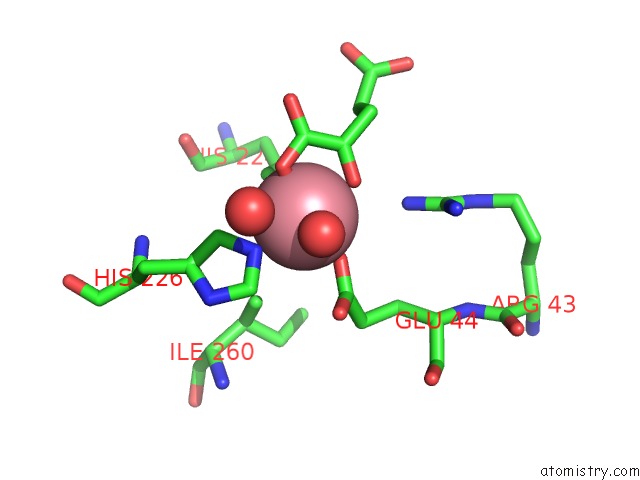
Mono view
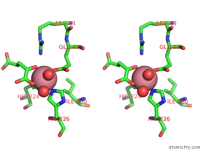
Stereo pair view
Mono view
Stereo pair view
A full contact list of Cobalt with other atoms in the Co binding
site number 1 of Homocitrate Synthase LYS4 Bound to 2-Og within 5.0Å range:
|
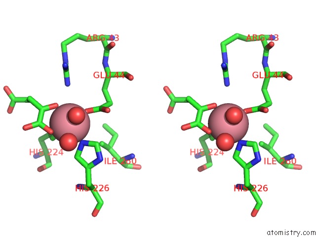
Cobalt binding site 2 out of 2 in 3ivu
Go back to
Cobalt binding site 2 out
of 2 in the Homocitrate Synthase LYS4 Bound to 2-Og

Mono view
Stereo pair view
Mono view
Stereo pair view
A full contact list of Cobalt with other atoms in the Co binding
site number 2 of Homocitrate Synthase LYS4 Bound to 2-Og within 5.0Å range:
|
Reference:
S.L.Bulfer,
E.M.Scott,
J.F.Couture,
L.Pillus,
R.C.Trievel.
Crystal Structure and Functional Analysis of Homocitrate Synthase, An Essential Enzyme in Lysine Biosynthesis. J.Biol.Chem. V. 284 35769 2009.
ISSN: ISSN 0021-9258
PubMed: 19776021
DOI: 10.1074/JBC.M109.046821
Page generated: Tue Jul 30 16:12:09 2024
ISSN: ISSN 0021-9258
PubMed: 19776021
DOI: 10.1074/JBC.M109.046821
Last articles
Zn in 9J0NZn in 9J0O
Zn in 9J0P
Zn in 9FJX
Zn in 9EKB
Zn in 9C0F
Zn in 9CAH
Zn in 9CH0
Zn in 9CH3
Zn in 9CH1