Cobalt »
PDB 3igz-3mcr »
3kok »
Cobalt in PDB 3kok: Crystal Structure of Cobalt (II) Human Carbonic Anhydrase II at pH 8.5
Enzymatic activity of Crystal Structure of Cobalt (II) Human Carbonic Anhydrase II at pH 8.5
All present enzymatic activity of Crystal Structure of Cobalt (II) Human Carbonic Anhydrase II at pH 8.5:
4.2.1.1;
4.2.1.1;
Protein crystallography data
The structure of Crystal Structure of Cobalt (II) Human Carbonic Anhydrase II at pH 8.5, PDB code: 3kok
was solved by
B.S.Avvaru,
with X-Ray Crystallography technique. A brief refinement statistics is given in the table below:
| Resolution Low / High (Å) | 20.00 / 1.50 |
| Space group | P 1 21 1 |
| Cell size a, b, c (Å), α, β, γ (°) | 42.366, 41.159, 72.062, 90.00, 104.28, 90.00 |
| R / Rfree (%) | 22 / 22.7 |
Cobalt Binding Sites:
The binding sites of Cobalt atom in the Crystal Structure of Cobalt (II) Human Carbonic Anhydrase II at pH 8.5
(pdb code 3kok). This binding sites where shown within
5.0 Angstroms radius around Cobalt atom.
In total only one binding site of Cobalt was determined in the Crystal Structure of Cobalt (II) Human Carbonic Anhydrase II at pH 8.5, PDB code: 3kok:
In total only one binding site of Cobalt was determined in the Crystal Structure of Cobalt (II) Human Carbonic Anhydrase II at pH 8.5, PDB code: 3kok:
Cobalt binding site 1 out of 1 in 3kok
Go back to
Cobalt binding site 1 out
of 1 in the Crystal Structure of Cobalt (II) Human Carbonic Anhydrase II at pH 8.5
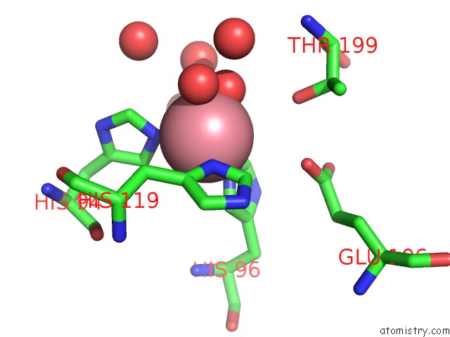
Mono view
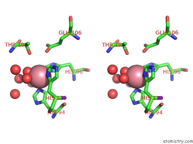
Stereo pair view
Mono view
Stereo pair view
A full contact list of Cobalt with other atoms in the Co binding
site number 1 of Crystal Structure of Cobalt (II) Human Carbonic Anhydrase II at pH 8.5 within 5.0Å range:
|
Reference:
B.S.Avvaru,
D.J.Arenas,
C.Tu,
D.B.Tanner,
R.Mckenna,
D.N.Silverman.
Comparison of Solution and Crystal Properties of Co(II)-Substituted Human Carbonic Anhydrase II. Arch.Biochem.Biophys. V. 502 53 2010.
ISSN: ISSN 0003-9861
PubMed: 20637176
DOI: 10.1016/J.ABB.2010.07.010
Page generated: Tue Jul 30 16:14:10 2024
ISSN: ISSN 0003-9861
PubMed: 20637176
DOI: 10.1016/J.ABB.2010.07.010
Last articles
Zn in 9MJ5Zn in 9HNW
Zn in 9G0L
Zn in 9FNE
Zn in 9DZN
Zn in 9E0I
Zn in 9D32
Zn in 9DAK
Zn in 8ZXC
Zn in 8ZUF