Cobalt »
PDB 3igz-3mcr »
3krk »
Cobalt in PDB 3krk: X-Ray Crystal Structure of Arachidonic Acid Bound in the Cyclooxygenase Channel of L531F Murine Cox-2
Enzymatic activity of X-Ray Crystal Structure of Arachidonic Acid Bound in the Cyclooxygenase Channel of L531F Murine Cox-2
All present enzymatic activity of X-Ray Crystal Structure of Arachidonic Acid Bound in the Cyclooxygenase Channel of L531F Murine Cox-2:
1.14.99.1;
1.14.99.1;
Protein crystallography data
The structure of X-Ray Crystal Structure of Arachidonic Acid Bound in the Cyclooxygenase Channel of L531F Murine Cox-2, PDB code: 3krk
was solved by
A.J.Vecchio,
D.M.Simmons,
M.G.Malkowski,
with X-Ray Crystallography technique. A brief refinement statistics is given in the table below:
| Resolution Low / High (Å) | 19.92 / 2.40 |
| Space group | I 2 2 2 |
| Cell size a, b, c (Å), α, β, γ (°) | 120.790, 133.040, 180.654, 90.00, 90.00, 90.00 |
| R / Rfree (%) | 17.9 / 22.8 |
Cobalt Binding Sites:
The binding sites of Cobalt atom in the X-Ray Crystal Structure of Arachidonic Acid Bound in the Cyclooxygenase Channel of L531F Murine Cox-2
(pdb code 3krk). This binding sites where shown within
5.0 Angstroms radius around Cobalt atom.
In total 2 binding sites of Cobalt where determined in the X-Ray Crystal Structure of Arachidonic Acid Bound in the Cyclooxygenase Channel of L531F Murine Cox-2, PDB code: 3krk:
Jump to Cobalt binding site number: 1; 2;
In total 2 binding sites of Cobalt where determined in the X-Ray Crystal Structure of Arachidonic Acid Bound in the Cyclooxygenase Channel of L531F Murine Cox-2, PDB code: 3krk:
Jump to Cobalt binding site number: 1; 2;
Cobalt binding site 1 out of 2 in 3krk
Go back to
Cobalt binding site 1 out
of 2 in the X-Ray Crystal Structure of Arachidonic Acid Bound in the Cyclooxygenase Channel of L531F Murine Cox-2
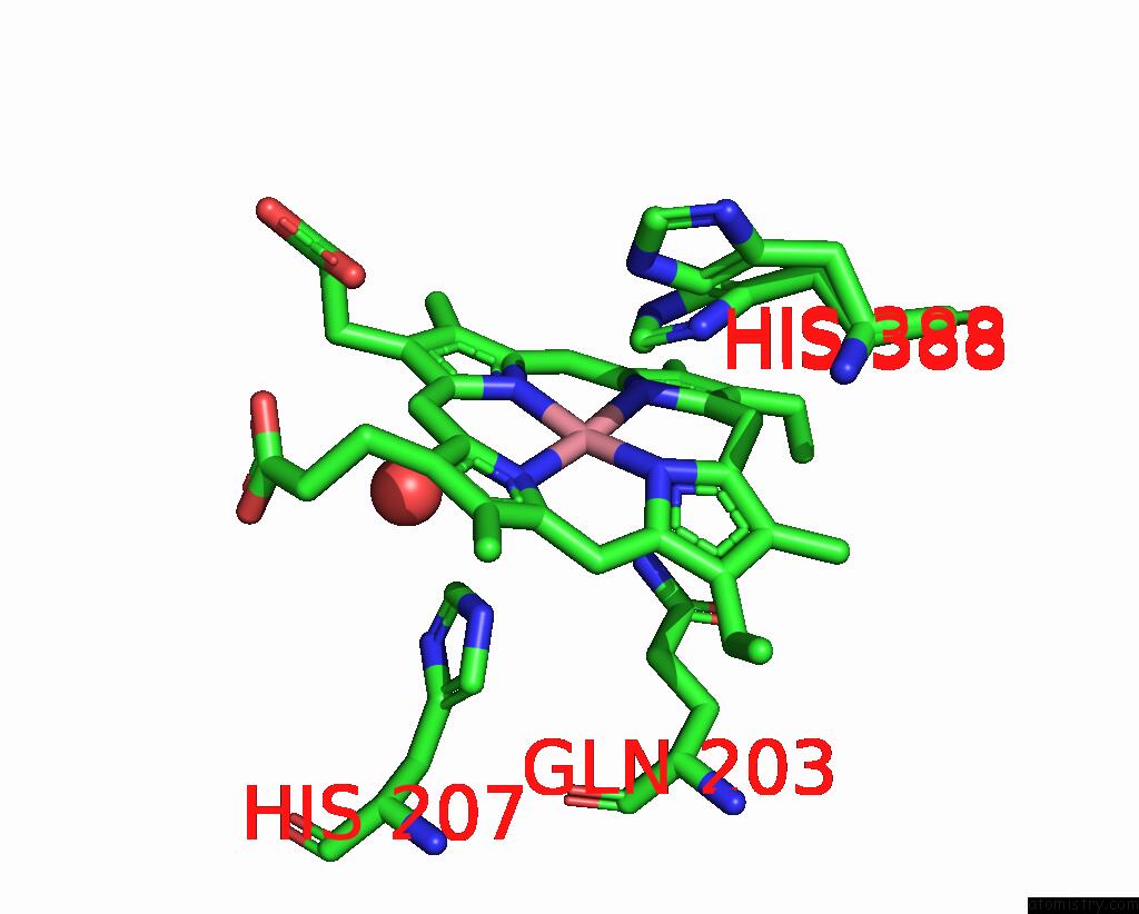
Mono view
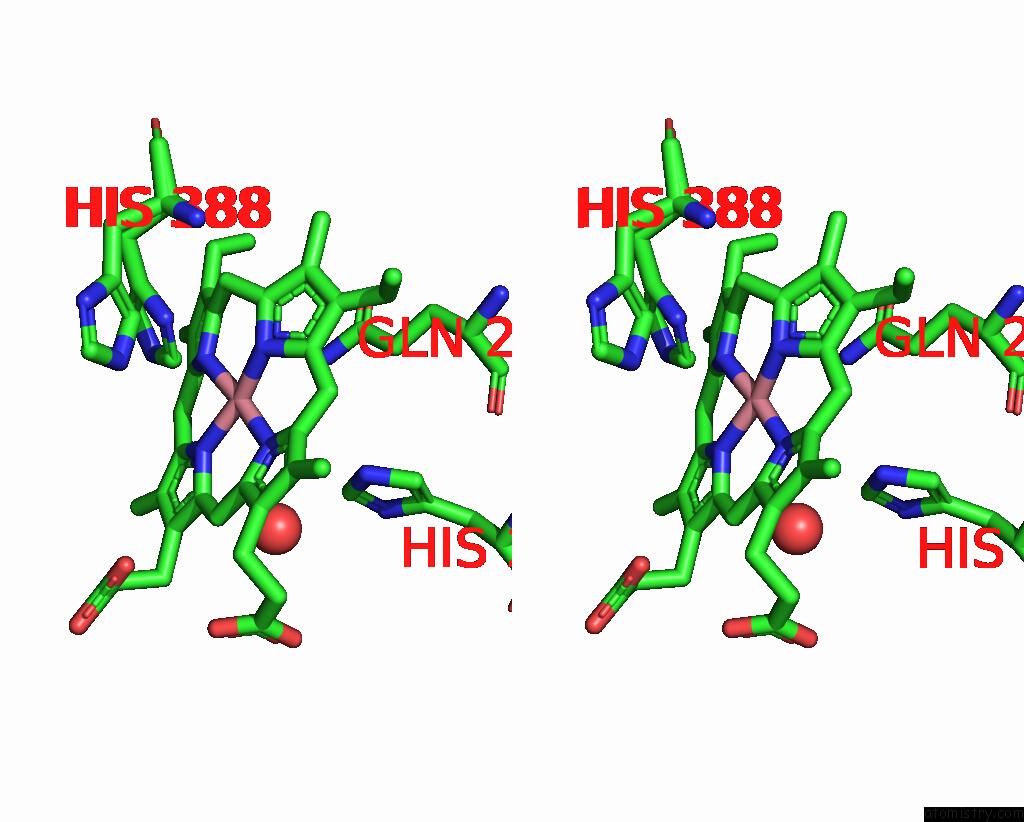
Stereo pair view
Mono view
Stereo pair view
A full contact list of Cobalt with other atoms in the Co binding
site number 1 of X-Ray Crystal Structure of Arachidonic Acid Bound in the Cyclooxygenase Channel of L531F Murine Cox-2 within 5.0Å range:
|
Cobalt binding site 2 out of 2 in 3krk
Go back to
Cobalt binding site 2 out
of 2 in the X-Ray Crystal Structure of Arachidonic Acid Bound in the Cyclooxygenase Channel of L531F Murine Cox-2
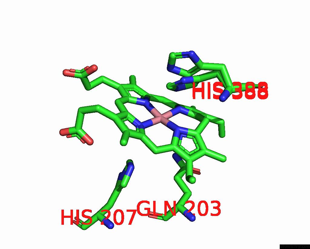
Mono view
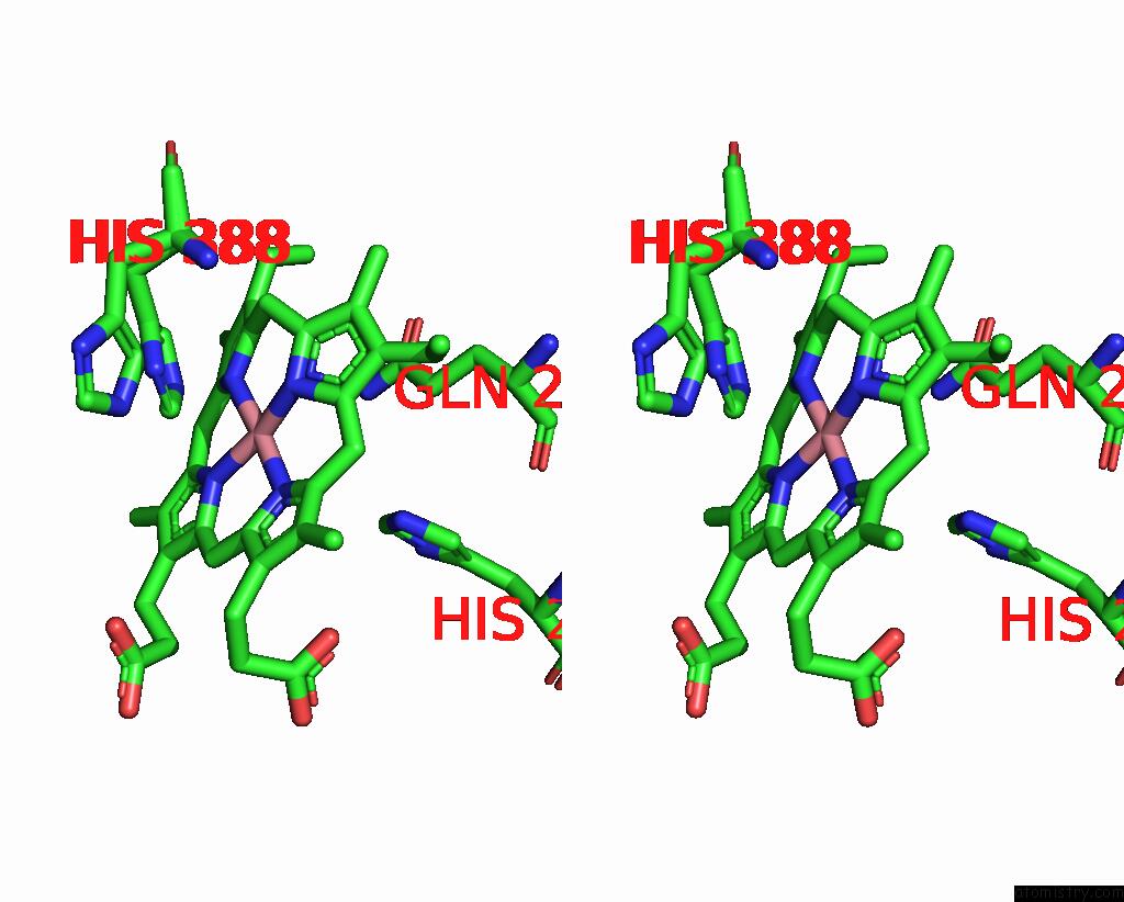
Stereo pair view
Mono view
Stereo pair view
A full contact list of Cobalt with other atoms in the Co binding
site number 2 of X-Ray Crystal Structure of Arachidonic Acid Bound in the Cyclooxygenase Channel of L531F Murine Cox-2 within 5.0Å range:
|
Reference:
A.J.Vecchio,
D.M.Simmons,
M.G.Malkowski.
Structural Basis of Fatty Acid Substrate Binding to Cyclooxygenase-2. J.Biol.Chem. V. 285 22152 2010.
ISSN: ISSN 0021-9258
PubMed: 20463020
DOI: 10.1074/JBC.M110.119867
Page generated: Tue Jul 30 16:17:14 2024
ISSN: ISSN 0021-9258
PubMed: 20463020
DOI: 10.1074/JBC.M110.119867
Last articles
Zn in 9MJ5Zn in 9HNW
Zn in 9G0L
Zn in 9FNE
Zn in 9DZN
Zn in 9E0I
Zn in 9D32
Zn in 9DAK
Zn in 8ZXC
Zn in 8ZUF