Cobalt »
PDB 3igz-3mcr »
3kyo »
Cobalt in PDB 3kyo: Crystal Structure of Hla-G Presenting Klpaqfyil Peptide
Protein crystallography data
The structure of Crystal Structure of Hla-G Presenting Klpaqfyil Peptide, PDB code: 3kyo
was solved by
N.G.Walpole,
J.Rossjohn,
C.S.Clements,
with X-Ray Crystallography technique. A brief refinement statistics is given in the table below:
| Resolution Low / High (Å) | 43.00 / 1.70 |
| Space group | P 1 21 1 |
| Cell size a, b, c (Å), α, β, γ (°) | 58.606, 85.982, 111.570, 90.00, 95.61, 90.00 |
| R / Rfree (%) | 18.2 / 22.6 |
Cobalt Binding Sites:
The binding sites of Cobalt atom in the Crystal Structure of Hla-G Presenting Klpaqfyil Peptide
(pdb code 3kyo). This binding sites where shown within
5.0 Angstroms radius around Cobalt atom.
In total 2 binding sites of Cobalt where determined in the Crystal Structure of Hla-G Presenting Klpaqfyil Peptide, PDB code: 3kyo:
Jump to Cobalt binding site number: 1; 2;
In total 2 binding sites of Cobalt where determined in the Crystal Structure of Hla-G Presenting Klpaqfyil Peptide, PDB code: 3kyo:
Jump to Cobalt binding site number: 1; 2;
Cobalt binding site 1 out of 2 in 3kyo
Go back to
Cobalt binding site 1 out
of 2 in the Crystal Structure of Hla-G Presenting Klpaqfyil Peptide
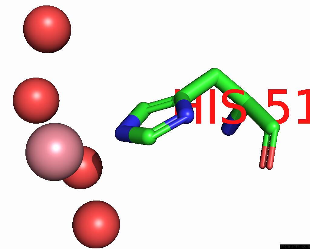
Mono view
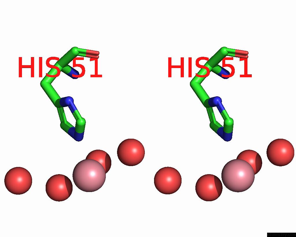
Stereo pair view
Mono view
Stereo pair view
A full contact list of Cobalt with other atoms in the Co binding
site number 1 of Crystal Structure of Hla-G Presenting Klpaqfyil Peptide within 5.0Å range:
|
Cobalt binding site 2 out of 2 in 3kyo
Go back to
Cobalt binding site 2 out
of 2 in the Crystal Structure of Hla-G Presenting Klpaqfyil Peptide
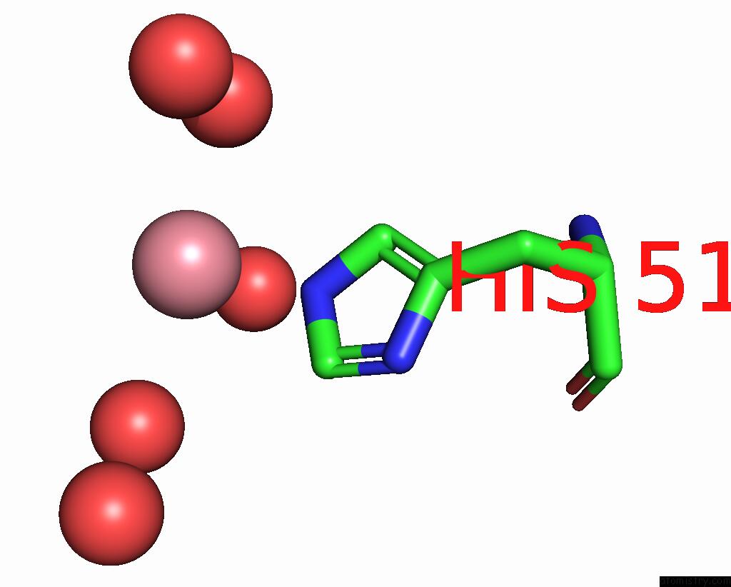
Mono view
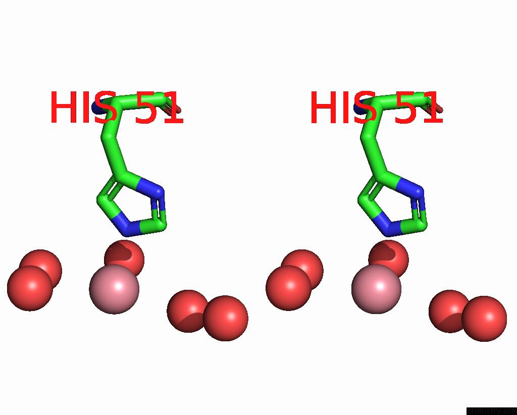
Stereo pair view
Mono view
Stereo pair view
A full contact list of Cobalt with other atoms in the Co binding
site number 2 of Crystal Structure of Hla-G Presenting Klpaqfyil Peptide within 5.0Å range:
|
Reference:
N.G.Walpole,
L.Kjer-Nielsen,
L.Kostenko,
J.Mccluskey,
A.G.Brooks,
J.Rossjohn,
C.S.Clements.
The Structure and Stability of the Monomorphic Hla-G Are Influenced By the Nature of the Bound Peptide J.Mol.Biol. V. 397 467 2010.
ISSN: ISSN 0022-2836
PubMed: 20122941
DOI: 10.1016/J.JMB.2010.01.052
Page generated: Tue Jul 30 16:17:45 2024
ISSN: ISSN 0022-2836
PubMed: 20122941
DOI: 10.1016/J.JMB.2010.01.052
Last articles
Zn in 9MJ5Zn in 9HNW
Zn in 9G0L
Zn in 9FNE
Zn in 9DZN
Zn in 9E0I
Zn in 9D32
Zn in 9DAK
Zn in 8ZXC
Zn in 8ZUF