Cobalt »
PDB 3igz-3mcr »
3l48 »
Cobalt in PDB 3l48: Crystal Structure of the C-Terminal Domain of the Papc Usher
Protein crystallography data
The structure of Crystal Structure of the C-Terminal Domain of the Papc Usher, PDB code: 3l48
was solved by
B.A.Ford,
S.J.Hultgren,
with X-Ray Crystallography technique. A brief refinement statistics is given in the table below:
| Resolution Low / High (Å) | 45.14 / 2.10 |
| Space group | P 42 21 2 |
| Cell size a, b, c (Å), α, β, γ (°) | 100.936, 100.936, 89.540, 90.00, 90.00, 90.00 |
| R / Rfree (%) | 20 / 23 |
Cobalt Binding Sites:
The binding sites of Cobalt atom in the Crystal Structure of the C-Terminal Domain of the Papc Usher
(pdb code 3l48). This binding sites where shown within
5.0 Angstroms radius around Cobalt atom.
In total 2 binding sites of Cobalt where determined in the Crystal Structure of the C-Terminal Domain of the Papc Usher, PDB code: 3l48:
Jump to Cobalt binding site number: 1; 2;
In total 2 binding sites of Cobalt where determined in the Crystal Structure of the C-Terminal Domain of the Papc Usher, PDB code: 3l48:
Jump to Cobalt binding site number: 1; 2;
Cobalt binding site 1 out of 2 in 3l48
Go back to
Cobalt binding site 1 out
of 2 in the Crystal Structure of the C-Terminal Domain of the Papc Usher
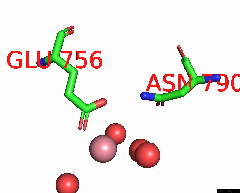
Mono view
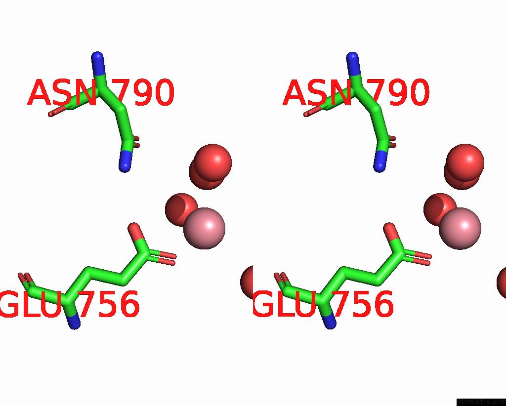
Stereo pair view
Mono view
Stereo pair view
A full contact list of Cobalt with other atoms in the Co binding
site number 1 of Crystal Structure of the C-Terminal Domain of the Papc Usher within 5.0Å range:
|
Cobalt binding site 2 out of 2 in 3l48
Go back to
Cobalt binding site 2 out
of 2 in the Crystal Structure of the C-Terminal Domain of the Papc Usher

Mono view
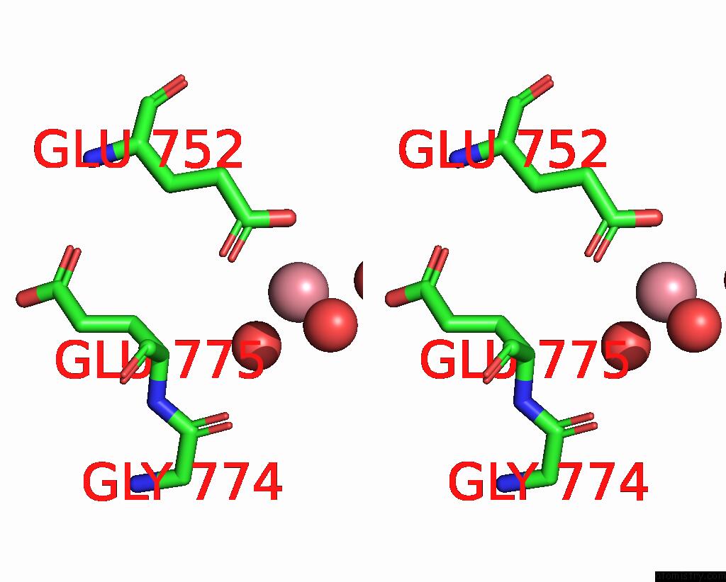
Stereo pair view
Mono view
Stereo pair view
A full contact list of Cobalt with other atoms in the Co binding
site number 2 of Crystal Structure of the C-Terminal Domain of the Papc Usher within 5.0Å range:
|
Reference:
B.Ford,
A.T.Rego,
T.J.Ragan,
J.Pinkner,
K.Dodson,
P.C.Driscoll,
S.Hultgren,
G.Waksman.
Structural Homology Between the C-Terminal Domain of the Papc Usher and Its Plug. J.Bacteriol. V. 192 1824 2010.
ISSN: ISSN 0021-9193
PubMed: 20118254
DOI: 10.1128/JB.01677-09
Page generated: Tue Jul 30 16:17:47 2024
ISSN: ISSN 0021-9193
PubMed: 20118254
DOI: 10.1128/JB.01677-09
Last articles
Zn in 9MJ5Zn in 9HNW
Zn in 9G0L
Zn in 9FNE
Zn in 9DZN
Zn in 9E0I
Zn in 9D32
Zn in 9DAK
Zn in 8ZXC
Zn in 8ZUF