Cobalt »
PDB 3igz-3mcr »
3m8d »
Cobalt in PDB 3m8d: Crystal Structure of Spin-Labeled Btub V10R1 with Bound Calcium and Cyanocobalamin
Protein crystallography data
The structure of Crystal Structure of Spin-Labeled Btub V10R1 with Bound Calcium and Cyanocobalamin, PDB code: 3m8d
was solved by
D.M.Freed,
P.S.Horanyi,
M.C.Wiener,
D.S.Cafiso,
with X-Ray Crystallography technique. A brief refinement statistics is given in the table below:
| Resolution Low / High (Å) | 44.04 / 2.44 |
| Space group | P 31 2 1 |
| Cell size a, b, c (Å), α, β, γ (°) | 82.051, 82.051, 224.457, 90.00, 90.00, 120.00 |
| R / Rfree (%) | 22.6 / 27.5 |
Other elements in 3m8d:
The structure of Crystal Structure of Spin-Labeled Btub V10R1 with Bound Calcium and Cyanocobalamin also contains other interesting chemical elements:
| Calcium | (Ca) | 3 atoms |
Cobalt Binding Sites:
The binding sites of Cobalt atom in the Crystal Structure of Spin-Labeled Btub V10R1 with Bound Calcium and Cyanocobalamin
(pdb code 3m8d). This binding sites where shown within
5.0 Angstroms radius around Cobalt atom.
In total only one binding site of Cobalt was determined in the Crystal Structure of Spin-Labeled Btub V10R1 with Bound Calcium and Cyanocobalamin, PDB code: 3m8d:
In total only one binding site of Cobalt was determined in the Crystal Structure of Spin-Labeled Btub V10R1 with Bound Calcium and Cyanocobalamin, PDB code: 3m8d:
Cobalt binding site 1 out of 1 in 3m8d
Go back to
Cobalt binding site 1 out
of 1 in the Crystal Structure of Spin-Labeled Btub V10R1 with Bound Calcium and Cyanocobalamin
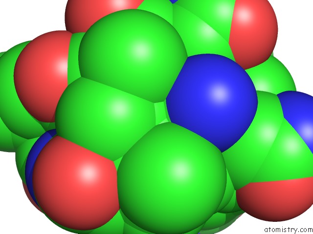
Mono view
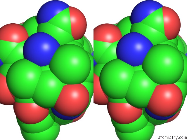
Stereo pair view
Mono view
Stereo pair view
A full contact list of Cobalt with other atoms in the Co binding
site number 1 of Crystal Structure of Spin-Labeled Btub V10R1 with Bound Calcium and Cyanocobalamin within 5.0Å range:
|
Reference:
D.M.Freed,
P.S.Horanyi,
M.C.Wiener,
D.S.Cafiso.
Conformational Exchange in A Membrane Transport Protein Is Altered in Protein Crystals. Biophys.J. V. 99 1604 2010.
ISSN: ISSN 0006-3495
PubMed: 20816073
DOI: 10.1016/J.BPJ.2010.06.026
Page generated: Tue Jul 30 16:18:54 2024
ISSN: ISSN 0006-3495
PubMed: 20816073
DOI: 10.1016/J.BPJ.2010.06.026
Last articles
Zn in 9J0NZn in 9J0O
Zn in 9J0P
Zn in 9FJX
Zn in 9EKB
Zn in 9C0F
Zn in 9CAH
Zn in 9CH0
Zn in 9CH3
Zn in 9CH1