Cobalt »
PDB 3mdl-3r61 »
3ngy »
Cobalt in PDB 3ngy: Crystal Structure of Rnase T (E92G Mutant)
Protein crystallography data
The structure of Crystal Structure of Rnase T (E92G Mutant), PDB code: 3ngy
was solved by
Y.-Y.Hsiao,
H.S.Yuan,
with X-Ray Crystallography technique. A brief refinement statistics is given in the table below:
| Resolution Low / High (Å) | 27.61 / 2.20 |
| Space group | P 21 21 21 |
| Cell size a, b, c (Å), α, β, γ (°) | 58.763, 107.718, 121.745, 90.00, 90.00, 90.00 |
| R / Rfree (%) | 17.5 / 22.4 |
Cobalt Binding Sites:
The binding sites of Cobalt atom in the Crystal Structure of Rnase T (E92G Mutant)
(pdb code 3ngy). This binding sites where shown within
5.0 Angstroms radius around Cobalt atom.
In total 3 binding sites of Cobalt where determined in the Crystal Structure of Rnase T (E92G Mutant), PDB code: 3ngy:
Jump to Cobalt binding site number: 1; 2; 3;
In total 3 binding sites of Cobalt where determined in the Crystal Structure of Rnase T (E92G Mutant), PDB code: 3ngy:
Jump to Cobalt binding site number: 1; 2; 3;
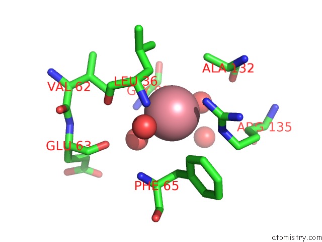
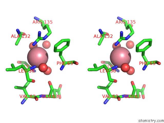
Cobalt binding site 1 out of 3 in 3ngy
Go back to
Cobalt binding site 1 out
of 3 in the Crystal Structure of Rnase T (E92G Mutant)
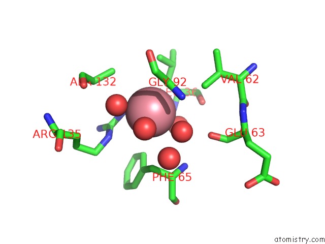
Mono view
Stereo pair view
Mono view
Stereo pair view
A full contact list of Cobalt with other atoms in the Co binding
site number 1 of Crystal Structure of Rnase T (E92G Mutant) within 5.0Å range:
|
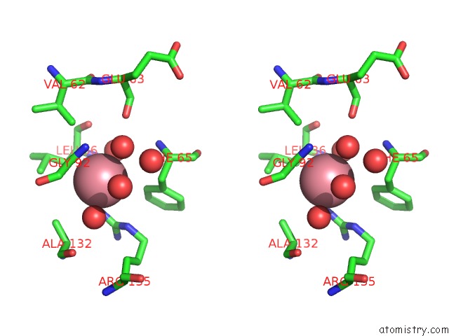
Cobalt binding site 2 out of 3 in 3ngy
Go back to
Cobalt binding site 2 out
of 3 in the Crystal Structure of Rnase T (E92G Mutant)
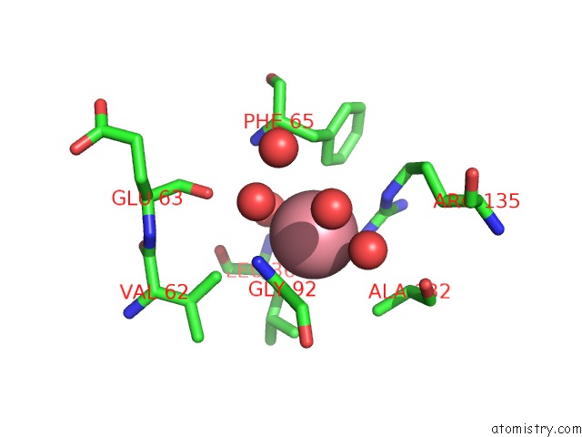
Mono view
Stereo pair view
Mono view
Stereo pair view
A full contact list of Cobalt with other atoms in the Co binding
site number 2 of Crystal Structure of Rnase T (E92G Mutant) within 5.0Å range:
|
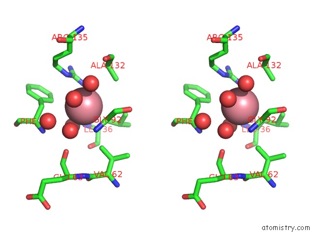
Cobalt binding site 3 out of 3 in 3ngy
Go back to
Cobalt binding site 3 out
of 3 in the Crystal Structure of Rnase T (E92G Mutant)
Mono view
Stereo pair view
Mono view
Stereo pair view
A full contact list of Cobalt with other atoms in the Co binding
site number 3 of Crystal Structure of Rnase T (E92G Mutant) within 5.0Å range:
|
Reference:
Y.-Y.Hsiao,
C.-C.Yang,
C.L.Lin,
J.L.J.Lin,
Y.Duh,
H.S.Yuan.
Structural Basis For Rna Trimming By Rnase T in Stable Rna 3'-End Maturation Nat.Chem.Biol. V. 7 236 2011.
ISSN: ISSN 1552-4450
PubMed: 21317904
DOI: 10.1038/NCHEMBIO.524
Page generated: Tue Jul 30 16:20:53 2024
ISSN: ISSN 1552-4450
PubMed: 21317904
DOI: 10.1038/NCHEMBIO.524
Last articles
Zn in 9MJ5Zn in 9HNW
Zn in 9G0L
Zn in 9FNE
Zn in 9DZN
Zn in 9E0I
Zn in 9D32
Zn in 9DAK
Zn in 8ZXC
Zn in 8ZUF