Cobalt »
PDB 3r86-3ura »
3sc0 »
Cobalt in PDB 3sc0: Crystal Structure of Mmachc (1-238), A Human B12 Processing Enzyme, Complexed with Methylcobalamin
Protein crystallography data
The structure of Crystal Structure of Mmachc (1-238), A Human B12 Processing Enzyme, Complexed with Methylcobalamin, PDB code: 3sc0
was solved by
M.Koutmos,
C.Gherasim,
J.L.Smith,
R.Banerjee,
with X-Ray Crystallography technique. A brief refinement statistics is given in the table below:
| Resolution Low / High (Å) | 40.29 / 1.95 |
| Space group | P 62 2 2 |
| Cell size a, b, c (Å), α, β, γ (°) | 105.955, 105.955, 84.253, 90.00, 90.00, 120.00 |
| R / Rfree (%) | 18.6 / 24.4 |
Cobalt Binding Sites:
The binding sites of Cobalt atom in the Crystal Structure of Mmachc (1-238), A Human B12 Processing Enzyme, Complexed with Methylcobalamin
(pdb code 3sc0). This binding sites where shown within
5.0 Angstroms radius around Cobalt atom.
In total only one binding site of Cobalt was determined in the Crystal Structure of Mmachc (1-238), A Human B12 Processing Enzyme, Complexed with Methylcobalamin, PDB code: 3sc0:
In total only one binding site of Cobalt was determined in the Crystal Structure of Mmachc (1-238), A Human B12 Processing Enzyme, Complexed with Methylcobalamin, PDB code: 3sc0:
Cobalt binding site 1 out of 1 in 3sc0
Go back to
Cobalt binding site 1 out
of 1 in the Crystal Structure of Mmachc (1-238), A Human B12 Processing Enzyme, Complexed with Methylcobalamin
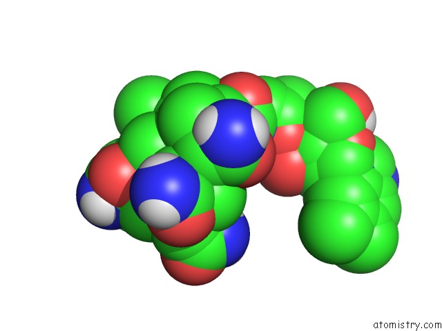
Mono view
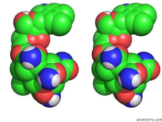
Stereo pair view
Mono view
Stereo pair view
A full contact list of Cobalt with other atoms in the Co binding
site number 1 of Crystal Structure of Mmachc (1-238), A Human B12 Processing Enzyme, Complexed with Methylcobalamin within 5.0Å range:
|
Reference:
M.Koutmos,
C.Gherasim,
J.L.Smith,
R.Banerjee.
Structural Basis of Multifunctionality in A Vitamin B12-Processing Enzyme. J.Biol.Chem. V. 286 29780 2011.
ISSN: ISSN 0021-9258
PubMed: 21697092
DOI: 10.1074/JBC.M111.261370
Page generated: Tue Jul 30 16:33:37 2024
ISSN: ISSN 0021-9258
PubMed: 21697092
DOI: 10.1074/JBC.M111.261370
Last articles
Zn in 9MJ5Zn in 9HNW
Zn in 9G0L
Zn in 9FNE
Zn in 9DZN
Zn in 9E0I
Zn in 9D32
Zn in 9DAK
Zn in 8ZXC
Zn in 8ZUF