Cobalt in PDB 3urn: Crystal Structure of Pte Mutant H254G/H257W/L303T/K185R/I274N/A80V/S61T with Cyclohexyl Methylphosphonate Inhibitor
Enzymatic activity of Crystal Structure of Pte Mutant H254G/H257W/L303T/K185R/I274N/A80V/S61T with Cyclohexyl Methylphosphonate Inhibitor
All present enzymatic activity of Crystal Structure of Pte Mutant H254G/H257W/L303T/K185R/I274N/A80V/S61T with Cyclohexyl Methylphosphonate Inhibitor:
3.1.8.1;
Protein crystallography data
The structure of Crystal Structure of Pte Mutant H254G/H257W/L303T/K185R/I274N/A80V/S61T with Cyclohexyl Methylphosphonate Inhibitor, PDB code: 3urn
was solved by
P.Tsai,
N.G.Fox,
Y.Li,
D.P.Barondeau,
F.M.Raushel,
with X-Ray Crystallography technique. A brief refinement statistics is given in the table below:
Resolution Low / High (Å)
|
35.81 /
1.95
|
Space group
|
P 21 21 2
|
Cell size a, b, c (Å), α, β, γ (°)
|
85.498,
86.103,
88.741,
90.00,
90.00,
90.00
|
R / Rfree (%)
|
22 /
24.7
|
Cobalt Binding Sites:
The binding sites of Cobalt atom in the Crystal Structure of Pte Mutant H254G/H257W/L303T/K185R/I274N/A80V/S61T with Cyclohexyl Methylphosphonate Inhibitor
(pdb code 3urn). This binding sites where shown within
5.0 Angstroms radius around Cobalt atom.
In total 4 binding sites of Cobalt where determined in the
Crystal Structure of Pte Mutant H254G/H257W/L303T/K185R/I274N/A80V/S61T with Cyclohexyl Methylphosphonate Inhibitor, PDB code: 3urn:
Jump to Cobalt binding site number:
1;
2;
3;
4;
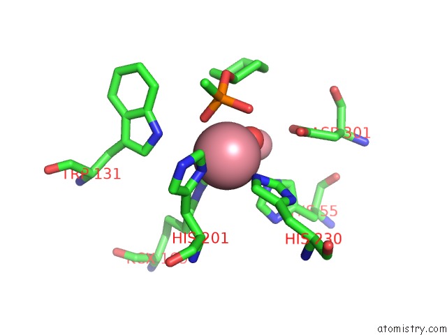
Cobalt binding site 1 out
of 4 in 3urn
Go back to
Cobalt Binding Sites List in 3urn
Cobalt binding site 1 out
of 4 in the Crystal Structure of Pte Mutant H254G/H257W/L303T/K185R/I274N/A80V/S61T with Cyclohexyl Methylphosphonate Inhibitor
Mono view
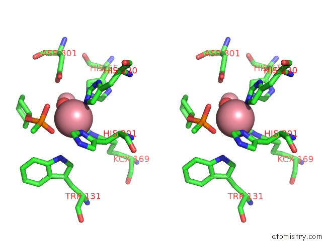
Stereo pair view
|
A full contact list of Cobalt with other atoms in the Co binding
site number 1 of Crystal Structure of Pte Mutant H254G/H257W/L303T/K185R/I274N/A80V/S61T with Cyclohexyl Methylphosphonate Inhibitor within 5.0Å range:
probe
|
atom
|
residue
|
distance (Å)
|
B
|
Occ
|
A:Co801
b:21.3
occ:1.00
|
NE2
|
A:HIS230
|
2.1
|
17.3
|
1.0
|
OQ2
|
A:KCX169
|
2.1
|
16.2
|
1.0
|
O
|
A:HOH1361
|
2.1
|
18.4
|
1.0
|
ND1
|
A:HIS201
|
2.2
|
19.1
|
1.0
|
OAC
|
A:QMP902
|
2.3
|
66.5
|
1.0
|
OAB
|
A:QMP902
|
2.9
|
66.5
|
1.0
|
CE1
|
A:HIS201
|
3.0
|
21.7
|
1.0
|
CD2
|
A:HIS230
|
3.0
|
18.7
|
1.0
|
CX
|
A:KCX169
|
3.0
|
19.2
|
1.0
|
CE1
|
A:HIS230
|
3.1
|
15.7
|
1.0
|
PAK
|
A:QMP902
|
3.1
|
67.6
|
1.0
|
CG
|
A:HIS201
|
3.3
|
20.6
|
1.0
|
OQ1
|
A:KCX169
|
3.3
|
19.0
|
1.0
|
CO
|
A:CO802
|
3.7
|
21.5
|
1.0
|
CB
|
A:HIS201
|
3.7
|
18.8
|
1.0
|
NE1
|
A:TRP131
|
4.0
|
24.8
|
1.0
|
ND1
|
A:HIS230
|
4.2
|
19.9
|
1.0
|
NE2
|
A:HIS55
|
4.2
|
21.1
|
1.0
|
CG
|
A:HIS230
|
4.2
|
19.2
|
1.0
|
NE2
|
A:HIS201
|
4.2
|
22.4
|
1.0
|
OD2
|
A:ASP301
|
4.2
|
19.5
|
1.0
|
NZ
|
A:KCX169
|
4.2
|
17.4
|
1.0
|
OAI
|
A:QMP902
|
4.3
|
67.5
|
1.0
|
CD2
|
A:HIS201
|
4.4
|
23.2
|
1.0
|
CAA
|
A:QMP902
|
4.4
|
67.5
|
1.0
|
CAJ
|
A:QMP902
|
4.4
|
67.7
|
1.0
|
CE1
|
A:HIS55
|
4.5
|
22.9
|
1.0
|
CA
|
A:HIS201
|
4.6
|
20.7
|
1.0
|
CD1
|
A:TRP131
|
4.7
|
23.6
|
1.0
|
CE
|
A:KCX169
|
4.7
|
17.4
|
1.0
|
OD1
|
A:ASP301
|
5.0
|
21.4
|
1.0
|
CG
|
A:ASP301
|
5.0
|
21.1
|
1.0
|
|
Cobalt binding site 2 out
of 4 in 3urn
Go back to
Cobalt Binding Sites List in 3urn
Cobalt binding site 2 out
of 4 in the Crystal Structure of Pte Mutant H254G/H257W/L303T/K185R/I274N/A80V/S61T with Cyclohexyl Methylphosphonate Inhibitor
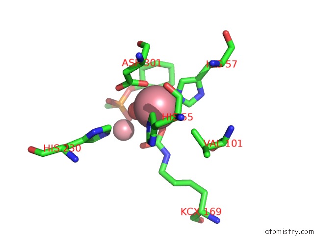
Mono view
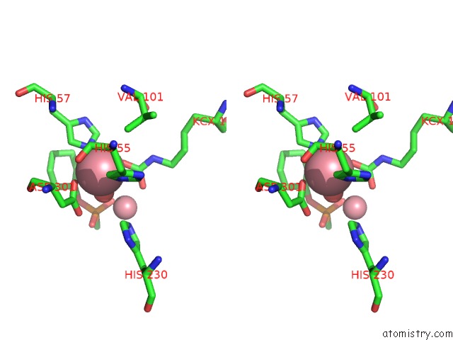
Stereo pair view
|
A full contact list of Cobalt with other atoms in the Co binding
site number 2 of Crystal Structure of Pte Mutant H254G/H257W/L303T/K185R/I274N/A80V/S61T with Cyclohexyl Methylphosphonate Inhibitor within 5.0Å range:
probe
|
atom
|
residue
|
distance (Å)
|
B
|
Occ
|
A:Co802
b:21.5
occ:1.00
|
O
|
A:HOH1361
|
1.8
|
18.4
|
1.0
|
NE2
|
A:HIS57
|
1.9
|
20.3
|
1.0
|
OQ1
|
A:KCX169
|
2.2
|
19.0
|
1.0
|
OD1
|
A:ASP301
|
2.3
|
21.4
|
1.0
|
NE2
|
A:HIS55
|
2.3
|
21.1
|
1.0
|
CE1
|
A:HIS57
|
2.9
|
21.3
|
1.0
|
CD2
|
A:HIS55
|
3.0
|
22.1
|
1.0
|
CD2
|
A:HIS57
|
3.0
|
20.7
|
1.0
|
CX
|
A:KCX169
|
3.1
|
19.2
|
1.0
|
CG
|
A:ASP301
|
3.1
|
21.1
|
1.0
|
CE1
|
A:HIS55
|
3.4
|
22.9
|
1.0
|
OD2
|
A:ASP301
|
3.4
|
19.5
|
1.0
|
OQ2
|
A:KCX169
|
3.6
|
16.2
|
1.0
|
CO
|
A:CO801
|
3.7
|
21.3
|
1.0
|
ND1
|
A:HIS57
|
4.0
|
19.0
|
1.0
|
CG2
|
A:VAL101
|
4.0
|
20.6
|
1.0
|
CG
|
A:HIS57
|
4.1
|
20.7
|
1.0
|
NZ
|
A:KCX169
|
4.1
|
17.4
|
1.0
|
CAJ
|
A:QMP902
|
4.1
|
67.7
|
1.0
|
CG
|
A:HIS55
|
4.2
|
22.9
|
1.0
|
CE1
|
A:HIS230
|
4.2
|
15.7
|
1.0
|
CAE
|
A:QMP902
|
4.3
|
67.6
|
1.0
|
ND1
|
A:HIS55
|
4.4
|
24.1
|
1.0
|
CAF
|
A:QMP902
|
4.4
|
67.8
|
1.0
|
NE2
|
A:HIS230
|
4.4
|
17.3
|
1.0
|
CB
|
A:ASP301
|
4.4
|
19.5
|
1.0
|
OAC
|
A:QMP902
|
4.6
|
66.5
|
1.0
|
CAG
|
A:QMP902
|
4.6
|
67.8
|
1.0
|
CAH
|
A:QMP902
|
4.7
|
68.2
|
1.0
|
OAB
|
A:QMP902
|
4.8
|
66.5
|
1.0
|
CA
|
A:ASP301
|
4.8
|
20.8
|
1.0
|
CAD
|
A:QMP902
|
4.9
|
67.6
|
1.0
|
|
Cobalt binding site 3 out
of 4 in 3urn
Go back to
Cobalt Binding Sites List in 3urn
Cobalt binding site 3 out
of 4 in the Crystal Structure of Pte Mutant H254G/H257W/L303T/K185R/I274N/A80V/S61T with Cyclohexyl Methylphosphonate Inhibitor
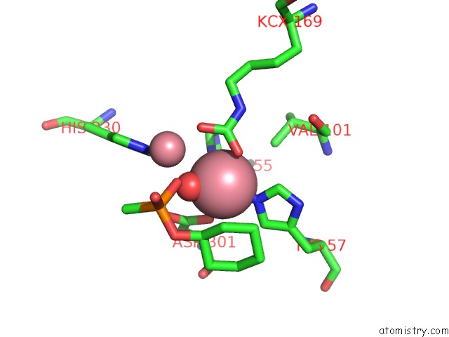
Mono view
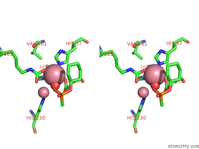
Stereo pair view
|
A full contact list of Cobalt with other atoms in the Co binding
site number 3 of Crystal Structure of Pte Mutant H254G/H257W/L303T/K185R/I274N/A80V/S61T with Cyclohexyl Methylphosphonate Inhibitor within 5.0Å range:
probe
|
atom
|
residue
|
distance (Å)
|
B
|
Occ
|
B:Co803
b:20.9
occ:1.00
|
O
|
B:HOH1368
|
1.9
|
13.6
|
1.0
|
NE2
|
B:HIS57
|
2.0
|
21.8
|
1.0
|
OQ2
|
B:KCX169
|
2.2
|
21.4
|
1.0
|
NE2
|
B:HIS55
|
2.3
|
22.8
|
1.0
|
OD1
|
B:ASP301
|
2.3
|
13.9
|
1.0
|
CD2
|
B:HIS55
|
3.0
|
21.1
|
1.0
|
CE1
|
B:HIS57
|
3.0
|
22.8
|
1.0
|
CX
|
B:KCX169
|
3.0
|
21.6
|
1.0
|
CD2
|
B:HIS57
|
3.0
|
22.4
|
1.0
|
CG
|
B:ASP301
|
3.1
|
15.4
|
1.0
|
OQ1
|
B:KCX169
|
3.3
|
25.0
|
1.0
|
CE1
|
B:HIS55
|
3.4
|
19.1
|
1.0
|
OD2
|
B:ASP301
|
3.4
|
19.8
|
1.0
|
OAC
|
B:QMP901
|
3.8
|
70.7
|
1.0
|
CO
|
B:CO804
|
3.8
|
24.9
|
1.0
|
CG2
|
B:VAL101
|
4.0
|
19.0
|
1.0
|
NZ
|
B:KCX169
|
4.1
|
20.0
|
1.0
|
ND1
|
B:HIS57
|
4.1
|
19.4
|
1.0
|
CG
|
B:HIS57
|
4.1
|
20.5
|
1.0
|
CAJ
|
B:QMP901
|
4.2
|
71.1
|
1.0
|
CG
|
B:HIS55
|
4.2
|
22.1
|
1.0
|
CE1
|
B:HIS230
|
4.2
|
23.1
|
1.0
|
ND1
|
B:HIS55
|
4.4
|
20.7
|
1.0
|
CB
|
B:ASP301
|
4.5
|
17.9
|
1.0
|
CAF
|
B:QMP901
|
4.5
|
71.1
|
1.0
|
NE2
|
B:HIS230
|
4.5
|
20.6
|
1.0
|
CAE
|
B:QMP901
|
4.7
|
70.7
|
1.0
|
CAH
|
B:QMP901
|
4.7
|
71.1
|
1.0
|
CAG
|
B:QMP901
|
4.9
|
70.9
|
1.0
|
CA
|
B:ASP301
|
4.9
|
20.8
|
1.0
|
|
Cobalt binding site 4 out
of 4 in 3urn
Go back to
Cobalt Binding Sites List in 3urn
Cobalt binding site 4 out
of 4 in the Crystal Structure of Pte Mutant H254G/H257W/L303T/K185R/I274N/A80V/S61T with Cyclohexyl Methylphosphonate Inhibitor
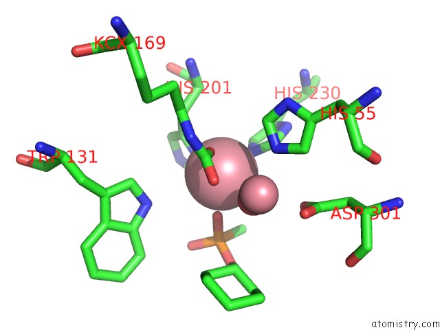
Mono view
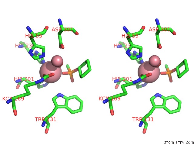
Stereo pair view
|
A full contact list of Cobalt with other atoms in the Co binding
site number 4 of Crystal Structure of Pte Mutant H254G/H257W/L303T/K185R/I274N/A80V/S61T with Cyclohexyl Methylphosphonate Inhibitor within 5.0Å range:
probe
|
atom
|
residue
|
distance (Å)
|
B
|
Occ
|
B:Co804
b:24.9
occ:1.00
|
NE2
|
B:HIS230
|
2.1
|
20.6
|
1.0
|
ND1
|
B:HIS201
|
2.1
|
29.1
|
1.0
|
OAC
|
B:QMP901
|
2.1
|
70.7
|
1.0
|
OQ1
|
B:KCX169
|
2.1
|
25.0
|
1.0
|
O
|
B:HOH1368
|
2.4
|
13.6
|
1.0
|
CE1
|
B:HIS201
|
2.9
|
30.8
|
1.0
|
CE1
|
B:HIS230
|
3.0
|
23.1
|
1.0
|
CD2
|
B:HIS230
|
3.1
|
23.4
|
1.0
|
CX
|
B:KCX169
|
3.2
|
21.6
|
1.0
|
CG
|
B:HIS201
|
3.2
|
30.1
|
1.0
|
PAK
|
B:QMP901
|
3.4
|
70.3
|
1.0
|
OQ2
|
B:KCX169
|
3.6
|
21.4
|
1.0
|
CB
|
B:HIS201
|
3.6
|
26.4
|
1.0
|
CAA
|
B:QMP901
|
3.7
|
70.4
|
1.0
|
CO
|
B:CO803
|
3.8
|
20.9
|
1.0
|
NE1
|
B:TRP131
|
4.0
|
27.0
|
1.0
|
NE2
|
B:HIS201
|
4.1
|
31.1
|
1.0
|
ND1
|
B:HIS230
|
4.1
|
21.8
|
1.0
|
OAB
|
B:QMP901
|
4.2
|
71.2
|
1.0
|
CG
|
B:HIS230
|
4.2
|
22.0
|
1.0
|
CD2
|
B:HIS201
|
4.2
|
29.2
|
1.0
|
NE2
|
B:HIS55
|
4.3
|
22.8
|
1.0
|
NZ
|
B:KCX169
|
4.3
|
20.0
|
1.0
|
OD2
|
B:ASP301
|
4.4
|
19.8
|
1.0
|
CE1
|
B:HIS55
|
4.5
|
19.1
|
1.0
|
CA
|
B:HIS201
|
4.5
|
25.3
|
1.0
|
OAI
|
B:QMP901
|
4.6
|
71.5
|
1.0
|
CD1
|
B:TRP131
|
4.6
|
26.4
|
1.0
|
CE
|
B:KCX169
|
4.7
|
19.8
|
1.0
|
CAJ
|
B:QMP901
|
4.7
|
71.1
|
1.0
|
|
Reference:
P.C.Tsai,
N.Fox,
A.N.Bigley,
S.P.Harvey,
D.P.Barondeau,
F.M.Raushel.
Enzymes For the Homeland Defense: Optimizing Phosphotriesterase For the Hydrolysis of Organophosphate Nerve Agents. Biochemistry V. 51 6463 2012.
ISSN: ISSN 0006-2960
PubMed: 22809162
DOI: 10.1021/BI300811T
Page generated: Tue Jul 30 16:46:10 2024
|