Cobalt »
PDB 3w72-4aq4 »
3wfu »
Cobalt in PDB 3wfu: Crystal Structure of Horse Heart Myoglobin Reconstituted with Cobalt(I) Tetradehydrocorrin
Protein crystallography data
The structure of Crystal Structure of Horse Heart Myoglobin Reconstituted with Cobalt(I) Tetradehydrocorrin, PDB code: 3wfu
was solved by
E.Mizohata,
Y.Morita,
K.Oohora,
K.Hirata,
J.Ohbayashi,
T.Inoue,
Y.Hisaeda,
T.Hayashi,
with X-Ray Crystallography technique. A brief refinement statistics is given in the table below:
| Resolution Low / High (Å) | 15.00 / 1.35 |
| Space group | P 1 21 1 |
| Cell size a, b, c (Å), α, β, γ (°) | 34.836, 28.720, 63.352, 90.00, 105.60, 90.00 |
| R / Rfree (%) | 14.3 / 19.7 |
Cobalt Binding Sites:
The binding sites of Cobalt atom in the Crystal Structure of Horse Heart Myoglobin Reconstituted with Cobalt(I) Tetradehydrocorrin
(pdb code 3wfu). This binding sites where shown within
5.0 Angstroms radius around Cobalt atom.
In total 2 binding sites of Cobalt where determined in the Crystal Structure of Horse Heart Myoglobin Reconstituted with Cobalt(I) Tetradehydrocorrin, PDB code: 3wfu:
Jump to Cobalt binding site number: 1; 2;
In total 2 binding sites of Cobalt where determined in the Crystal Structure of Horse Heart Myoglobin Reconstituted with Cobalt(I) Tetradehydrocorrin, PDB code: 3wfu:
Jump to Cobalt binding site number: 1; 2;
Cobalt binding site 1 out of 2 in 3wfu
Go back to
Cobalt binding site 1 out
of 2 in the Crystal Structure of Horse Heart Myoglobin Reconstituted with Cobalt(I) Tetradehydrocorrin
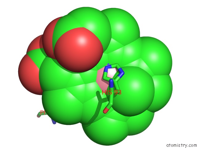
Mono view
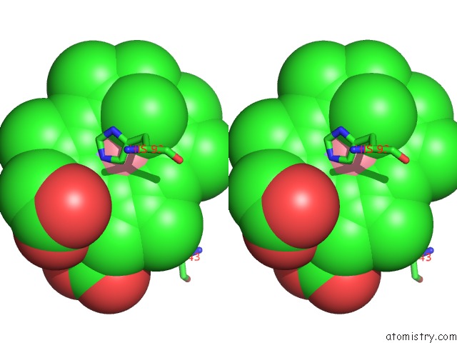
Stereo pair view
Mono view
Stereo pair view
A full contact list of Cobalt with other atoms in the Co binding
site number 1 of Crystal Structure of Horse Heart Myoglobin Reconstituted with Cobalt(I) Tetradehydrocorrin within 5.0Å range:
|
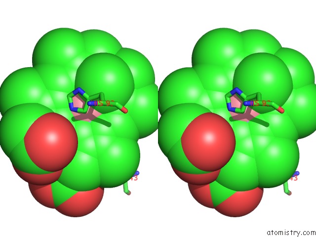
Cobalt binding site 2 out of 2 in 3wfu
Go back to
Cobalt binding site 2 out
of 2 in the Crystal Structure of Horse Heart Myoglobin Reconstituted with Cobalt(I) Tetradehydrocorrin

Mono view
Stereo pair view
Mono view
Stereo pair view
A full contact list of Cobalt with other atoms in the Co binding
site number 2 of Crystal Structure of Horse Heart Myoglobin Reconstituted with Cobalt(I) Tetradehydrocorrin within 5.0Å range:
|
Reference:
T.Hayashi,
Y.Morita,
E.Mizohata,
K.Oohora,
J.Ohbayashi,
T.Inoue,
Y.Hisaeda.
Co(II)/Co(I) Reduction-Induced Axial Histidine-Flipping in Myoglobin Reconstituted with A Cobalt Tetradehydrocorrin As A Methionine Synthase Model. Chem.Commun.(Camb.) V. 50 12560 2014.
ISSN: ISSN 1359-7345
PubMed: 25197974
DOI: 10.1039/C4CC05448B
Page generated: Sun Jul 13 19:36:14 2025
ISSN: ISSN 1359-7345
PubMed: 25197974
DOI: 10.1039/C4CC05448B
Last articles
Fe in 2YXOFe in 2YRS
Fe in 2YXC
Fe in 2YNM
Fe in 2YVJ
Fe in 2YP1
Fe in 2YU2
Fe in 2YU1
Fe in 2YQB
Fe in 2YOO