Cobalt »
PDB 4as7-4fck »
4cv7 »
Cobalt in PDB 4cv7: Crystal Structure of Rhodococcus Equi Vapb
Protein crystallography data
The structure of Crystal Structure of Rhodococcus Equi Vapb, PDB code: 4cv7
was solved by
C.Geerds,
H.H.Niemann,
with X-Ray Crystallography technique. A brief refinement statistics is given in the table below:
| Resolution Low / High (Å) | 50.00 / 1.40 |
| Space group | P 61 2 2 |
| Cell size a, b, c (Å), α, β, γ (°) | 83.820, 83.820, 49.130, 90.00, 90.00, 120.00 |
| R / Rfree (%) | 16.972 / 19.854 |
Cobalt Binding Sites:
The binding sites of Cobalt atom in the Crystal Structure of Rhodococcus Equi Vapb
(pdb code 4cv7). This binding sites where shown within
5.0 Angstroms radius around Cobalt atom.
In total 2 binding sites of Cobalt where determined in the Crystal Structure of Rhodococcus Equi Vapb, PDB code: 4cv7:
Jump to Cobalt binding site number: 1; 2;
In total 2 binding sites of Cobalt where determined in the Crystal Structure of Rhodococcus Equi Vapb, PDB code: 4cv7:
Jump to Cobalt binding site number: 1; 2;
Cobalt binding site 1 out of 2 in 4cv7
Go back to
Cobalt binding site 1 out
of 2 in the Crystal Structure of Rhodococcus Equi Vapb
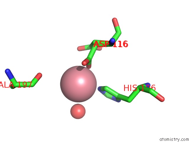
Mono view
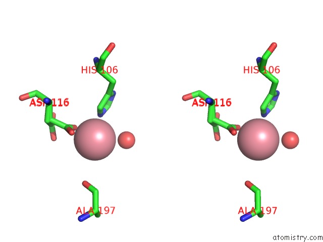
Stereo pair view
Mono view
Stereo pair view
A full contact list of Cobalt with other atoms in the Co binding
site number 1 of Crystal Structure of Rhodococcus Equi Vapb within 5.0Å range:
|
Cobalt binding site 2 out of 2 in 4cv7
Go back to
Cobalt binding site 2 out
of 2 in the Crystal Structure of Rhodococcus Equi Vapb
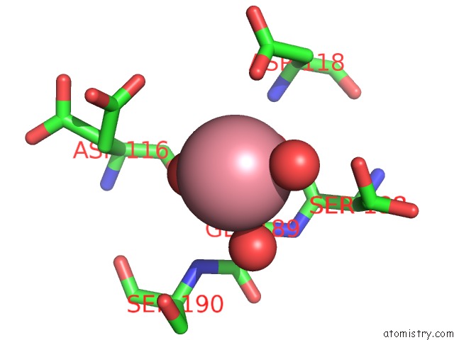
Mono view
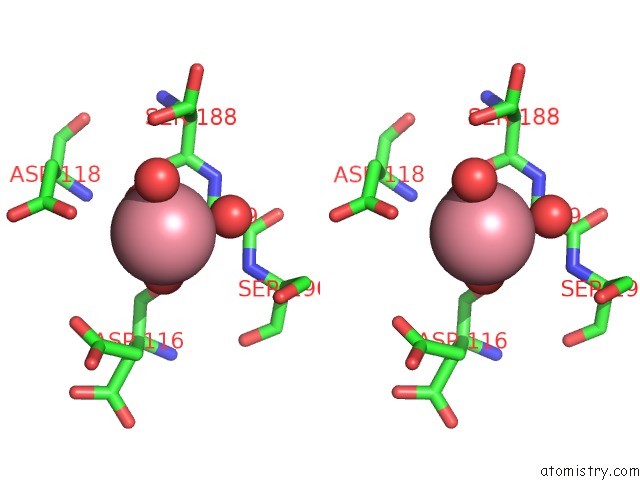
Stereo pair view
Mono view
Stereo pair view
A full contact list of Cobalt with other atoms in the Co binding
site number 2 of Crystal Structure of Rhodococcus Equi Vapb within 5.0Å range:
|
Reference:
C.Geerds,
J.Wohlmann,
A.Haas,
H.H.Niemann.
Structure of Rhodococcus Equi Virulence-Associated Protein B (Vapb) Reveals An Eight-Stranded Antiparallel [Beta]-Barrel Consisting of Two Greek-Key Motifs Acta Crystallogr.,Sect.F V. 70 866 2014.
ISSN: ISSN 1744-3091
PubMed: 25005079
DOI: 10.1107/S2053230X14009911
Page generated: Tue Jul 30 16:59:57 2024
ISSN: ISSN 1744-3091
PubMed: 25005079
DOI: 10.1107/S2053230X14009911
Last articles
Zn in 9JYWZn in 9IR4
Zn in 9IR3
Zn in 9GMX
Zn in 9GMW
Zn in 9JEJ
Zn in 9ERF
Zn in 9ERE
Zn in 9EGV
Zn in 9EGW