Cobalt »
PDB 4as7-4fck »
4dje »
Cobalt in PDB 4dje: Crystal Structure of Folate-Bound Corrinoid Iron-Sulfur Protein (Cfesp) in Complex with Its Methyltransferase (Metr), Co-Crystallized with Folate
Protein crystallography data
The structure of Crystal Structure of Folate-Bound Corrinoid Iron-Sulfur Protein (Cfesp) in Complex with Its Methyltransferase (Metr), Co-Crystallized with Folate, PDB code: 4dje
was solved by
Y.Kung,
C.L.Drennan,
with X-Ray Crystallography technique. A brief refinement statistics is given in the table below:
| Resolution Low / High (Å) | 48.46 / 3.50 |
| Space group | P 21 21 2 |
| Cell size a, b, c (Å), α, β, γ (°) | 136.419, 250.664, 82.427, 90.00, 90.00, 90.00 |
| R / Rfree (%) | 24 / 28.9 |
Other elements in 4dje:
The structure of Crystal Structure of Folate-Bound Corrinoid Iron-Sulfur Protein (Cfesp) in Complex with Its Methyltransferase (Metr), Co-Crystallized with Folate also contains other interesting chemical elements:
| Iron | (Fe) | 8 atoms |
| Calcium | (Ca) | 2 atoms |
Cobalt Binding Sites:
The binding sites of Cobalt atom in the Crystal Structure of Folate-Bound Corrinoid Iron-Sulfur Protein (Cfesp) in Complex with Its Methyltransferase (Metr), Co-Crystallized with Folate
(pdb code 4dje). This binding sites where shown within
5.0 Angstroms radius around Cobalt atom.
In total 2 binding sites of Cobalt where determined in the Crystal Structure of Folate-Bound Corrinoid Iron-Sulfur Protein (Cfesp) in Complex with Its Methyltransferase (Metr), Co-Crystallized with Folate, PDB code: 4dje:
Jump to Cobalt binding site number: 1; 2;
In total 2 binding sites of Cobalt where determined in the Crystal Structure of Folate-Bound Corrinoid Iron-Sulfur Protein (Cfesp) in Complex with Its Methyltransferase (Metr), Co-Crystallized with Folate, PDB code: 4dje:
Jump to Cobalt binding site number: 1; 2;
Cobalt binding site 1 out of 2 in 4dje
Go back to
Cobalt binding site 1 out
of 2 in the Crystal Structure of Folate-Bound Corrinoid Iron-Sulfur Protein (Cfesp) in Complex with Its Methyltransferase (Metr), Co-Crystallized with Folate
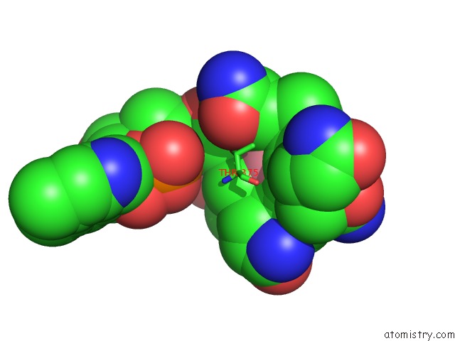
Mono view
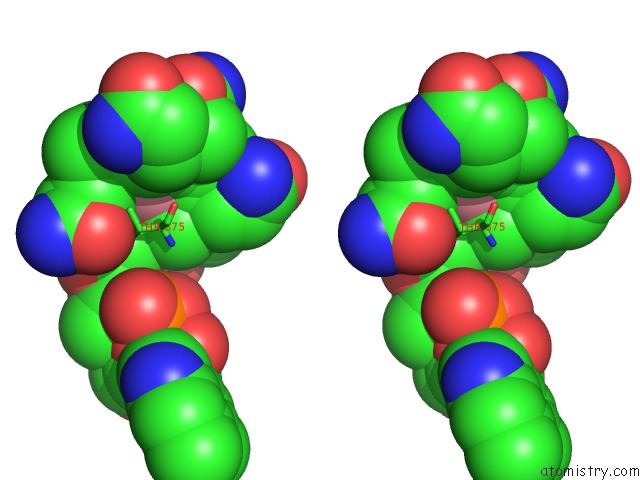
Stereo pair view
Mono view
Stereo pair view
A full contact list of Cobalt with other atoms in the Co binding
site number 1 of Crystal Structure of Folate-Bound Corrinoid Iron-Sulfur Protein (Cfesp) in Complex with Its Methyltransferase (Metr), Co-Crystallized with Folate within 5.0Å range:
|
Cobalt binding site 2 out of 2 in 4dje
Go back to
Cobalt binding site 2 out
of 2 in the Crystal Structure of Folate-Bound Corrinoid Iron-Sulfur Protein (Cfesp) in Complex with Its Methyltransferase (Metr), Co-Crystallized with Folate
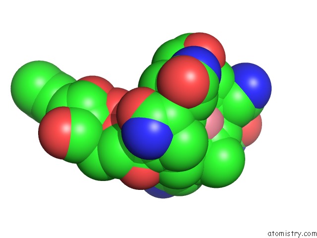
Mono view
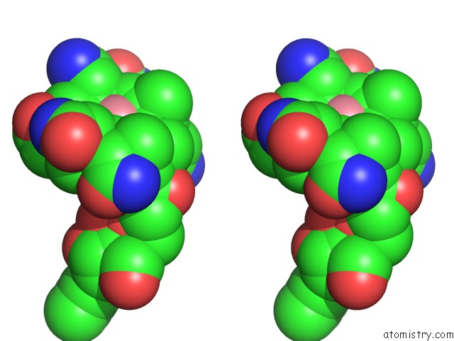
Stereo pair view
Mono view
Stereo pair view
A full contact list of Cobalt with other atoms in the Co binding
site number 2 of Crystal Structure of Folate-Bound Corrinoid Iron-Sulfur Protein (Cfesp) in Complex with Its Methyltransferase (Metr), Co-Crystallized with Folate within 5.0Å range:
|
Reference:
Y.Kung,
N.Ando,
T.I.Doukov,
L.C.Blasiak,
G.Bender,
J.Seravalli,
S.W.Ragsdale,
C.L.Drennan.
Visualizing Molecular Juggling Within A B12-Dependent Methyltransferase Complex. Nature V. 484 265 2012.
ISSN: ISSN 0028-0836
PubMed: 22419154
DOI: 10.1038/NATURE10916
Page generated: Tue Jul 30 17:01:49 2024
ISSN: ISSN 0028-0836
PubMed: 22419154
DOI: 10.1038/NATURE10916
Last articles
Zn in 9MJ5Zn in 9HNW
Zn in 9G0L
Zn in 9FNE
Zn in 9DZN
Zn in 9E0I
Zn in 9D32
Zn in 9DAK
Zn in 8ZXC
Zn in 8ZUF