Cobalt in PDB 4feo: Crystal Structure of the AU25A/A46G/C74U Mutant Xpt-Pbux Guanine Riboswitch Aptamer Domain in Complex with 2,6-Diaminopurine
Protein crystallography data
The structure of Crystal Structure of the AU25A/A46G/C74U Mutant Xpt-Pbux Guanine Riboswitch Aptamer Domain in Complex with 2,6-Diaminopurine, PDB code: 4feo
was solved by
C.D.Stoddard,
J.J.Trausch,
J.Widmann,
J.Marcano,
R.Knight,
R.T.Batey,
with X-Ray Crystallography technique. A brief refinement statistics is given in the table below:
Resolution Low / High (Å)
|
20.00 /
1.60
|
Space group
|
C 1 2 1
|
Cell size a, b, c (Å), α, β, γ (°)
|
132.910,
35.230,
41.460,
90.00,
90.36,
90.00
|
R / Rfree (%)
|
17.4 /
22.8
|
Cobalt Binding Sites:
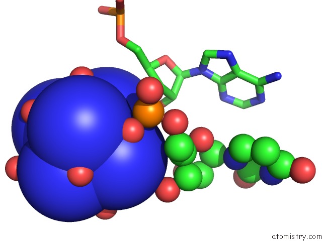
The binding sites of Cobalt atom in the Crystal Structure of the AU25A/A46G/C74U Mutant Xpt-Pbux Guanine Riboswitch Aptamer Domain in Complex with 2,6-Diaminopurine
(pdb code 4feo). This binding sites where shown within
5.0 Angstroms radius around Cobalt atom.
In total 9 binding sites of Cobalt where determined in the
Crystal Structure of the AU25A/A46G/C74U Mutant Xpt-Pbux Guanine Riboswitch Aptamer Domain in Complex with 2,6-Diaminopurine, PDB code: 4feo:
Jump to Cobalt binding site number:
1;
2;
3;
4;
5;
6;
7;
8;
9;
Cobalt binding site 1 out
of 9 in 4feo
Go back to
Cobalt Binding Sites List in 4feo
Cobalt binding site 1 out
of 9 in the Crystal Structure of the AU25A/A46G/C74U Mutant Xpt-Pbux Guanine Riboswitch Aptamer Domain in Complex with 2,6-Diaminopurine
Mono view
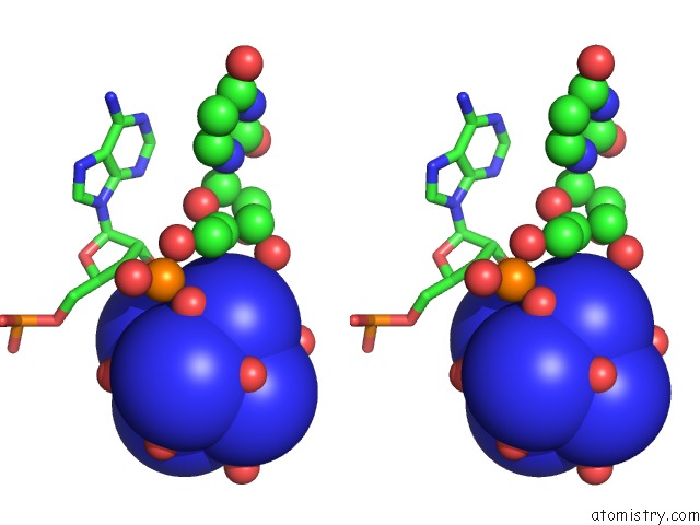
Stereo pair view
|
A full contact list of Cobalt with other atoms in the Co binding
site number 1 of Crystal Structure of the AU25A/A46G/C74U Mutant Xpt-Pbux Guanine Riboswitch Aptamer Domain in Complex with 2,6-Diaminopurine within 5.0Å range:
probe
|
atom
|
residue
|
distance (Å)
|
B
|
Occ
|
B:Co102
b:14.6
occ:1.00
|
CO
|
B:NCO102
|
0.0
|
14.6
|
1.0
|
N1
|
B:NCO102
|
2.0
|
15.6
|
1.0
|
N2
|
B:NCO102
|
2.0
|
15.3
|
1.0
|
N5
|
B:NCO102
|
2.0
|
12.8
|
1.0
|
N6
|
B:NCO102
|
2.0
|
14.7
|
1.0
|
N4
|
B:NCO102
|
2.0
|
15.0
|
1.0
|
N3
|
B:NCO102
|
2.0
|
15.1
|
1.0
|
O
|
B:HOH566
|
3.7
|
22.2
|
1.0
|
O
|
B:HOH311
|
3.8
|
15.7
|
1.0
|
O
|
B:HOH361
|
3.9
|
16.7
|
1.0
|
O
|
B:HOH422
|
3.9
|
22.9
|
1.0
|
O
|
B:HOH509
|
4.0
|
18.1
|
1.0
|
OP1
|
B:U67
|
4.1
|
11.1
|
1.0
|
O
|
B:HOH493
|
4.3
|
31.1
|
1.0
|
O3'
|
B:A66
|
4.3
|
11.0
|
1.0
|
O
|
B:HOH456
|
4.4
|
16.6
|
1.0
|
P
|
B:U67
|
4.9
|
10.0
|
1.0
|
O2'
|
B:A66
|
4.9
|
11.7
|
1.0
|
|
Cobalt binding site 2 out
of 9 in 4feo
Go back to
Cobalt Binding Sites List in 4feo
Cobalt binding site 2 out
of 9 in the Crystal Structure of the AU25A/A46G/C74U Mutant Xpt-Pbux Guanine Riboswitch Aptamer Domain in Complex with 2,6-Diaminopurine
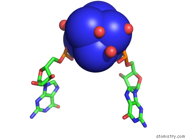
Mono view
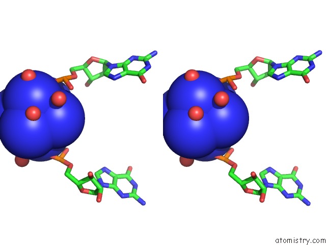
Stereo pair view
|
A full contact list of Cobalt with other atoms in the Co binding
site number 2 of Crystal Structure of the AU25A/A46G/C74U Mutant Xpt-Pbux Guanine Riboswitch Aptamer Domain in Complex with 2,6-Diaminopurine within 5.0Å range:
probe
|
atom
|
residue
|
distance (Å)
|
B
|
Occ
|
B:Co103
b:29.0
occ:1.00
|
CO
|
B:NCO103
|
0.0
|
29.0
|
1.0
|
N3
|
B:NCO103
|
2.0
|
25.4
|
1.0
|
N2
|
B:NCO103
|
2.0
|
25.9
|
1.0
|
N4
|
B:NCO103
|
2.0
|
26.2
|
1.0
|
N6
|
B:NCO103
|
2.0
|
25.1
|
1.0
|
N5
|
B:NCO103
|
2.0
|
26.5
|
1.0
|
N1
|
B:NCO103
|
2.0
|
26.2
|
1.0
|
O
|
B:HOH331
|
3.8
|
18.2
|
1.0
|
OP1
|
B:G38
|
3.8
|
10.7
|
1.0
|
O
|
B:HOH697
|
3.8
|
26.8
|
1.0
|
O
|
B:HOH746
|
3.9
|
37.1
|
1.0
|
O
|
B:HOH271
|
4.1
|
23.9
|
1.0
|
OP1
|
B:G56
|
4.1
|
20.9
|
1.0
|
O
|
B:HOH575
|
4.1
|
25.6
|
1.0
|
O
|
B:HOH712
|
4.7
|
42.4
|
1.0
|
O
|
B:HOH313
|
4.8
|
45.5
|
1.0
|
P
|
B:G38
|
4.9
|
9.6
|
1.0
|
OP2
|
B:G56
|
4.9
|
19.1
|
1.0
|
O
|
B:HOH465
|
4.9
|
32.3
|
1.0
|
OP2
|
B:G38
|
5.0
|
12.1
|
1.0
|
O
|
B:HOH268
|
5.0
|
23.6
|
1.0
|
|
Cobalt binding site 3 out
of 9 in 4feo
Go back to
Cobalt Binding Sites List in 4feo
Cobalt binding site 3 out
of 9 in the Crystal Structure of the AU25A/A46G/C74U Mutant Xpt-Pbux Guanine Riboswitch Aptamer Domain in Complex with 2,6-Diaminopurine
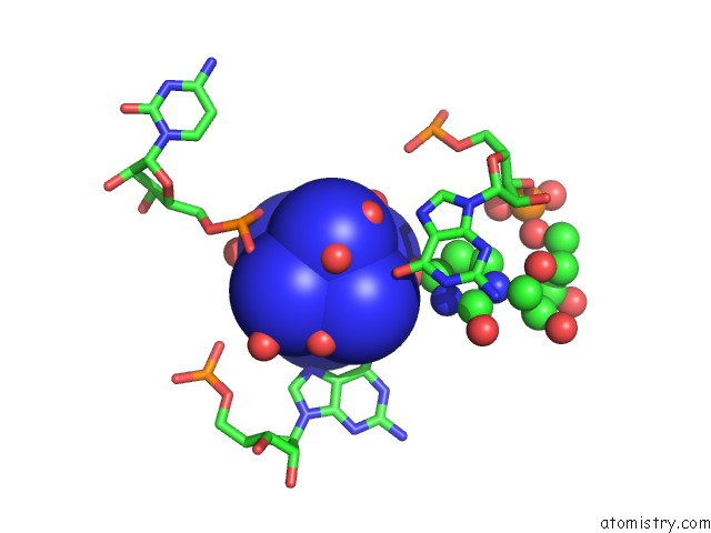
Mono view
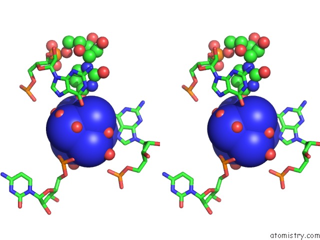
Stereo pair view
|
A full contact list of Cobalt with other atoms in the Co binding
site number 3 of Crystal Structure of the AU25A/A46G/C74U Mutant Xpt-Pbux Guanine Riboswitch Aptamer Domain in Complex with 2,6-Diaminopurine within 5.0Å range:
probe
|
atom
|
residue
|
distance (Å)
|
B
|
Occ
|
B:Co104
b:19.1
occ:1.00
|
CO
|
B:NCO104
|
0.0
|
19.1
|
1.0
|
N6
|
B:NCO104
|
1.9
|
20.2
|
1.0
|
N3
|
B:NCO104
|
2.0
|
16.0
|
1.0
|
N1
|
B:NCO104
|
2.0
|
19.2
|
1.0
|
N4
|
B:NCO104
|
2.0
|
21.9
|
1.0
|
N2
|
B:NCO104
|
2.0
|
20.3
|
1.0
|
N5
|
B:NCO104
|
2.0
|
19.8
|
1.0
|
O
|
B:HOH450
|
3.9
|
20.6
|
1.0
|
O
|
B:HOH306
|
4.0
|
22.2
|
1.0
|
O4
|
B:U69
|
4.0
|
10.3
|
1.0
|
O
|
B:HOH347
|
4.0
|
24.4
|
1.0
|
O6
|
B:G56
|
4.1
|
15.3
|
1.0
|
O
|
B:HOH250
|
4.1
|
22.0
|
1.0
|
O
|
B:HOH714
|
4.1
|
54.8
|
1.0
|
O
|
B:HOH620
|
4.2
|
39.8
|
1.0
|
O
|
B:HOH224
|
4.2
|
17.0
|
1.0
|
O
|
B:HOH728
|
4.4
|
56.5
|
1.0
|
O
|
B:HOH706
|
4.4
|
31.0
|
1.0
|
OP2
|
B:C39
|
4.4
|
13.7
|
1.0
|
N7
|
B:G56
|
4.5
|
14.5
|
1.0
|
OP1
|
B:C39
|
4.5
|
11.9
|
1.0
|
O
|
B:HOH651
|
4.5
|
0.7
|
1.0
|
O6
|
B:G68
|
4.8
|
12.5
|
1.0
|
C6
|
B:G56
|
4.9
|
15.0
|
1.0
|
O
|
B:HOH511
|
5.0
|
28.7
|
1.0
|
|
Cobalt binding site 4 out
of 9 in 4feo
Go back to
Cobalt Binding Sites List in 4feo
Cobalt binding site 4 out
of 9 in the Crystal Structure of the AU25A/A46G/C74U Mutant Xpt-Pbux Guanine Riboswitch Aptamer Domain in Complex with 2,6-Diaminopurine
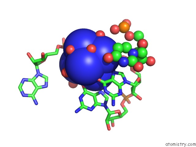
Mono view
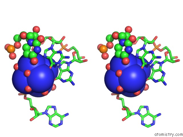
Stereo pair view
|
A full contact list of Cobalt with other atoms in the Co binding
site number 4 of Crystal Structure of the AU25A/A46G/C74U Mutant Xpt-Pbux Guanine Riboswitch Aptamer Domain in Complex with 2,6-Diaminopurine within 5.0Å range:
probe
|
atom
|
residue
|
distance (Å)
|
B
|
Occ
|
B:Co105
b:12.6
occ:1.00
|
CO
|
B:NCO105
|
0.0
|
12.6
|
1.0
|
N1
|
B:NCO105
|
2.0
|
10.2
|
1.0
|
N2
|
B:NCO105
|
2.0
|
11.1
|
1.0
|
N6
|
B:NCO105
|
2.0
|
10.8
|
1.0
|
N5
|
B:NCO105
|
2.0
|
12.3
|
1.0
|
N3
|
B:NCO105
|
2.0
|
11.2
|
1.0
|
N4
|
B:NCO105
|
2.0
|
8.8
|
1.0
|
OP2
|
B:A66
|
3.8
|
14.1
|
1.0
|
O
|
B:HOH214
|
3.8
|
13.6
|
1.0
|
O6
|
B:G32
|
3.9
|
11.6
|
1.0
|
O
|
B:HOH276
|
4.0
|
14.8
|
1.0
|
O6
|
B:G31
|
4.0
|
13.6
|
1.0
|
O
|
B:HOH231
|
4.1
|
13.6
|
1.0
|
O
|
B:HOH442
|
4.2
|
19.1
|
1.0
|
O
|
B:HOH305
|
4.2
|
12.9
|
1.0
|
O
|
B:HOH212
|
4.2
|
10.3
|
1.0
|
O
|
B:HOH254
|
4.2
|
13.6
|
1.0
|
O
|
B:HOH227
|
4.2
|
13.7
|
1.0
|
N7
|
B:G31
|
4.3
|
10.6
|
1.0
|
C6
|
B:G31
|
4.8
|
11.0
|
1.0
|
O
|
B:HOH206
|
4.9
|
12.5
|
1.0
|
O4
|
B:U30
|
4.9
|
11.9
|
1.0
|
C6
|
B:G32
|
4.9
|
11.8
|
1.0
|
C5
|
B:G31
|
5.0
|
10.1
|
1.0
|
|
Cobalt binding site 5 out
of 9 in 4feo
Go back to
Cobalt Binding Sites List in 4feo
Cobalt binding site 5 out
of 9 in the Crystal Structure of the AU25A/A46G/C74U Mutant Xpt-Pbux Guanine Riboswitch Aptamer Domain in Complex with 2,6-Diaminopurine
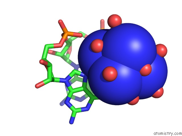
Mono view
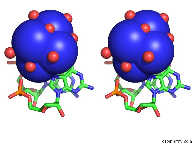
Stereo pair view
|
A full contact list of Cobalt with other atoms in the Co binding
site number 5 of Crystal Structure of the AU25A/A46G/C74U Mutant Xpt-Pbux Guanine Riboswitch Aptamer Domain in Complex with 2,6-Diaminopurine within 5.0Å range:
probe
|
atom
|
residue
|
distance (Å)
|
B
|
Occ
|
B:Co106
b:33.3
occ:1.00
|
CO
|
B:NCO106
|
0.0
|
33.3
|
1.0
|
N5
|
B:NCO106
|
2.0
|
34.1
|
1.0
|
N4
|
B:NCO106
|
2.0
|
34.9
|
1.0
|
N1
|
B:NCO106
|
2.0
|
33.6
|
1.0
|
N6
|
B:NCO106
|
2.0
|
33.2
|
1.0
|
N2
|
B:NCO106
|
2.0
|
33.4
|
1.0
|
N3
|
B:NCO106
|
2.0
|
31.5
|
1.0
|
O
|
B:HOH461
|
3.7
|
23.2
|
1.0
|
O
|
B:HOH316
|
3.8
|
27.5
|
1.0
|
OP2
|
B:G37
|
4.0
|
13.8
|
1.0
|
O
|
B:HOH258
|
4.0
|
18.6
|
1.0
|
O
|
B:HOH364
|
4.1
|
24.2
|
1.0
|
O6
|
B:G38
|
4.1
|
12.1
|
1.0
|
N7
|
B:G38
|
4.2
|
10.8
|
1.0
|
O
|
B:HOH851
|
4.3
|
41.6
|
1.0
|
O
|
B:HOH379
|
4.4
|
25.1
|
1.0
|
N7
|
B:G37
|
4.5
|
12.2
|
1.0
|
O
|
B:HOH569
|
4.6
|
22.9
|
1.0
|
O
|
B:HOH407
|
4.7
|
25.3
|
1.0
|
C6
|
B:G38
|
4.9
|
10.8
|
1.0
|
O
|
B:HOH339
|
4.9
|
23.9
|
1.0
|
C5
|
B:G38
|
5.0
|
10.6
|
1.0
|
O
|
B:HOH431
|
5.0
|
39.9
|
1.0
|
|
Cobalt binding site 6 out
of 9 in 4feo
Go back to
Cobalt Binding Sites List in 4feo
Cobalt binding site 6 out
of 9 in the Crystal Structure of the AU25A/A46G/C74U Mutant Xpt-Pbux Guanine Riboswitch Aptamer Domain in Complex with 2,6-Diaminopurine
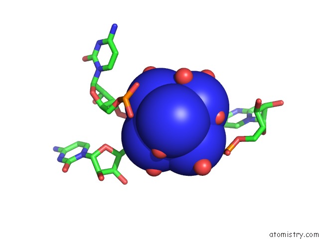
Mono view
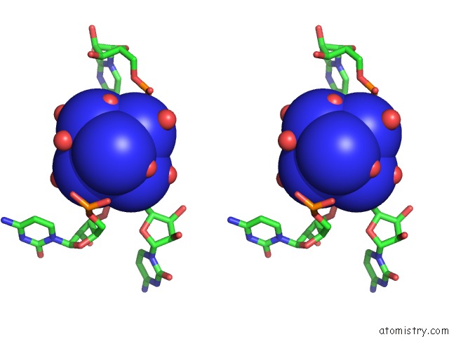
Stereo pair view
|
A full contact list of Cobalt with other atoms in the Co binding
site number 6 of Crystal Structure of the AU25A/A46G/C74U Mutant Xpt-Pbux Guanine Riboswitch Aptamer Domain in Complex with 2,6-Diaminopurine within 5.0Å range:
probe
|
atom
|
residue
|
distance (Å)
|
B
|
Occ
|
B:Co107
b:81.3
occ:1.00
|
CO
|
B:NCO107
|
0.0
|
81.3
|
1.0
|
N4
|
B:NCO107
|
2.0
|
81.0
|
1.0
|
N6
|
B:NCO107
|
2.0
|
81.1
|
1.0
|
N5
|
B:NCO107
|
2.0
|
81.2
|
1.0
|
N1
|
B:NCO107
|
2.0
|
81.1
|
1.0
|
N2
|
B:NCO107
|
2.0
|
80.9
|
1.0
|
N3
|
B:NCO107
|
2.0
|
81.0
|
1.0
|
O
|
B:HOH341
|
3.2
|
26.6
|
1.0
|
O
|
B:HOH409
|
3.5
|
23.0
|
1.0
|
O
|
B:HOH721
|
3.8
|
0.8
|
1.0
|
O
|
B:HOH615
|
3.8
|
55.4
|
1.0
|
O
|
B:HOH577
|
3.9
|
33.4
|
1.0
|
OP1
|
B:C54
|
4.0
|
24.4
|
1.0
|
OP2
|
B:C41
|
4.2
|
14.1
|
1.0
|
O
|
B:HOH640
|
4.3
|
41.4
|
1.0
|
OP1
|
B:C53
|
4.3
|
20.2
|
1.0
|
O
|
B:HOH613
|
4.3
|
43.9
|
1.0
|
O5'
|
B:C54
|
4.4
|
19.2
|
1.0
|
O
|
B:HOH590
|
4.6
|
35.5
|
1.0
|
OP2
|
B:C53
|
4.8
|
21.3
|
1.0
|
O
|
B:HOH348
|
4.8
|
33.4
|
1.0
|
O
|
B:HOH516
|
4.8
|
34.7
|
1.0
|
P
|
B:C54
|
4.9
|
21.8
|
1.0
|
P
|
B:C53
|
4.9
|
21.0
|
1.0
|
O
|
B:HOH228
|
5.0
|
15.5
|
1.0
|
|
Cobalt binding site 7 out
of 9 in 4feo
Go back to
Cobalt Binding Sites List in 4feo
Cobalt binding site 7 out
of 9 in the Crystal Structure of the AU25A/A46G/C74U Mutant Xpt-Pbux Guanine Riboswitch Aptamer Domain in Complex with 2,6-Diaminopurine
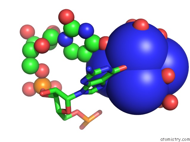
Mono view
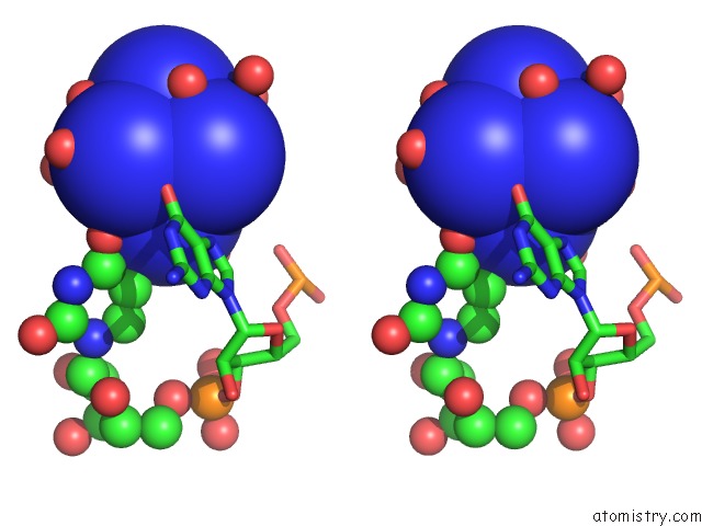
Stereo pair view
|
A full contact list of Cobalt with other atoms in the Co binding
site number 7 of Crystal Structure of the AU25A/A46G/C74U Mutant Xpt-Pbux Guanine Riboswitch Aptamer Domain in Complex with 2,6-Diaminopurine within 5.0Å range:
probe
|
atom
|
residue
|
distance (Å)
|
B
|
Occ
|
B:Co108
b:24.9
occ:1.00
|
CO
|
B:NCO108
|
0.0
|
24.9
|
1.0
|
N4
|
B:NCO108
|
2.0
|
24.6
|
1.0
|
N5
|
B:NCO108
|
2.0
|
24.6
|
1.0
|
N6
|
B:NCO108
|
2.0
|
24.5
|
1.0
|
N2
|
B:NCO108
|
2.0
|
24.7
|
1.0
|
N1
|
B:NCO108
|
2.0
|
23.8
|
1.0
|
N3
|
B:NCO108
|
2.0
|
25.1
|
1.0
|
O
|
B:HOH675
|
3.9
|
51.8
|
1.0
|
O
|
B:HOH392
|
3.9
|
30.1
|
1.0
|
O
|
B:HOH528
|
3.9
|
36.3
|
1.0
|
O
|
B:HOH366
|
4.0
|
36.6
|
1.0
|
O
|
B:HOH520
|
4.0
|
55.5
|
1.0
|
O
|
B:HOH595
|
4.1
|
33.1
|
1.0
|
O
|
B:HOH226
|
4.1
|
22.7
|
1.0
|
O6
|
B:G46
|
4.1
|
15.1
|
1.0
|
O4
|
B:U47
|
4.2
|
21.3
|
1.0
|
O
|
B:HOH328
|
4.3
|
24.5
|
1.0
|
N7
|
B:G46
|
4.3
|
18.2
|
1.0
|
O
|
B:HOH290
|
4.5
|
30.1
|
1.0
|
O
|
B:HOH255
|
4.9
|
33.8
|
1.0
|
C6
|
B:G46
|
4.9
|
17.8
|
1.0
|
O
|
B:HOH462
|
5.0
|
27.0
|
1.0
|
|
Cobalt binding site 8 out
of 9 in 4feo
Go back to
Cobalt Binding Sites List in 4feo
Cobalt binding site 8 out
of 9 in the Crystal Structure of the AU25A/A46G/C74U Mutant Xpt-Pbux Guanine Riboswitch Aptamer Domain in Complex with 2,6-Diaminopurine
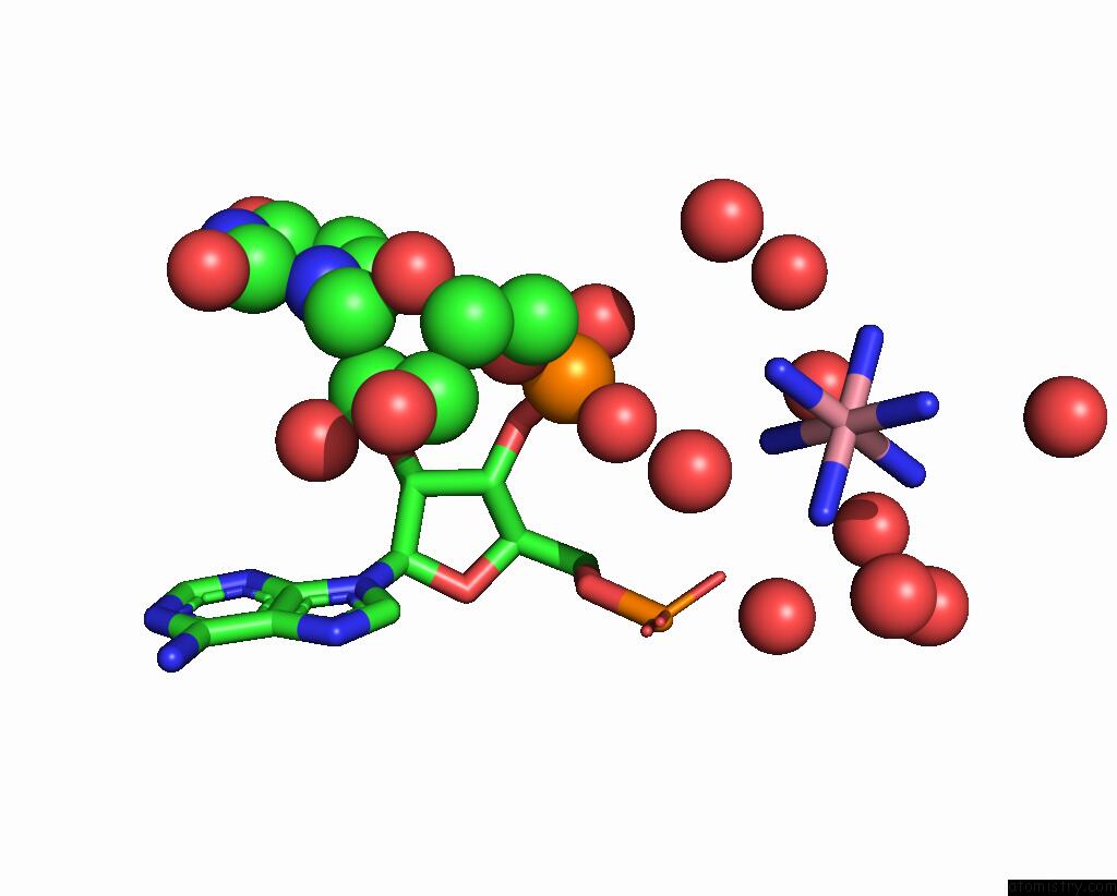
Mono view
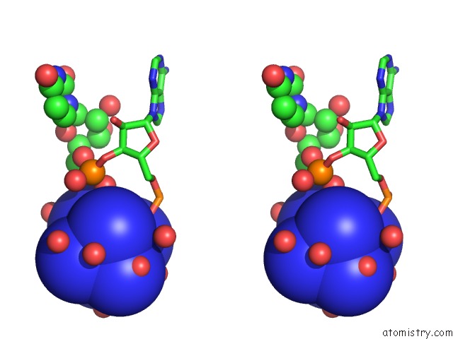
Stereo pair view
|
A full contact list of Cobalt with other atoms in the Co binding
site number 8 of Crystal Structure of the AU25A/A46G/C74U Mutant Xpt-Pbux Guanine Riboswitch Aptamer Domain in Complex with 2,6-Diaminopurine within 5.0Å range:
probe
|
atom
|
residue
|
distance (Å)
|
B
|
Occ
|
B:Co109
b:0.7
occ:1.00
|
CO
|
B:NCO109
|
0.0
|
0.7
|
1.0
|
N4
|
B:NCO109
|
2.0
|
0.5
|
1.0
|
N6
|
B:NCO109
|
2.0
|
0.5
|
1.0
|
N5
|
B:NCO109
|
2.0
|
0.5
|
1.0
|
N2
|
B:NCO109
|
2.0
|
0.5
|
1.0
|
N1
|
B:NCO109
|
2.0
|
0.6
|
1.0
|
N3
|
B:NCO109
|
2.0
|
0.5
|
1.0
|
O
|
B:HOH416
|
3.7
|
31.7
|
1.0
|
O
|
B:HOH859
|
3.9
|
64.8
|
1.0
|
OP1
|
B:A21
|
4.0
|
21.9
|
1.0
|
O
|
B:HOH624
|
4.1
|
53.5
|
1.0
|
O
|
B:HOH855
|
4.1
|
65.5
|
1.0
|
OP2
|
B:U22
|
4.4
|
18.0
|
1.0
|
O
|
B:HOH676
|
4.4
|
53.8
|
1.0
|
O
|
B:HOH669
|
4.5
|
42.1
|
1.0
|
O
|
B:HOH685
|
4.7
|
47.8
|
1.0
|
O
|
B:HOH663
|
4.7
|
51.0
|
1.0
|
O
|
B:HOH593
|
5.0
|
32.8
|
1.0
|
|
Cobalt binding site 9 out
of 9 in 4feo
Go back to
Cobalt Binding Sites List in 4feo
Cobalt binding site 9 out
of 9 in the Crystal Structure of the AU25A/A46G/C74U Mutant Xpt-Pbux Guanine Riboswitch Aptamer Domain in Complex with 2,6-Diaminopurine
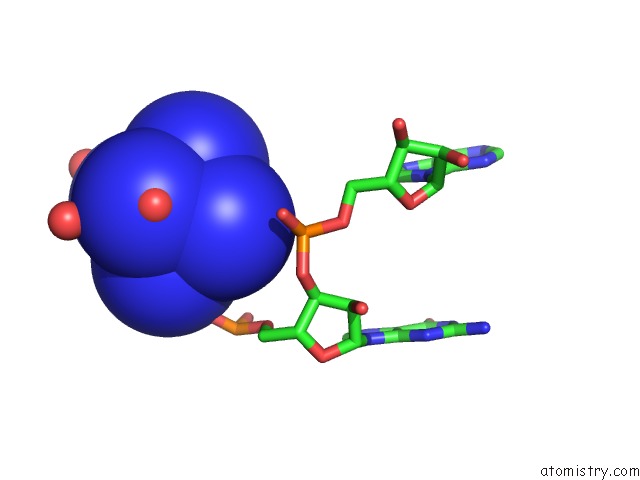
Mono view
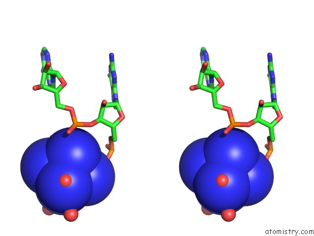
Stereo pair view
|
A full contact list of Cobalt with other atoms in the Co binding
site number 9 of Crystal Structure of the AU25A/A46G/C74U Mutant Xpt-Pbux Guanine Riboswitch Aptamer Domain in Complex with 2,6-Diaminopurine within 5.0Å range:
probe
|
atom
|
residue
|
distance (Å)
|
B
|
Occ
|
B:Co110
b:29.9
occ:0.64
|
CO
|
B:NCO110
|
0.0
|
29.9
|
0.6
|
N4
|
B:NCO110
|
2.0
|
28.1
|
0.6
|
N2
|
B:NCO110
|
2.0
|
29.8
|
0.6
|
N5
|
B:NCO110
|
2.0
|
30.1
|
0.6
|
N6
|
B:NCO110
|
2.0
|
29.1
|
0.6
|
N1
|
B:NCO110
|
2.0
|
30.4
|
0.6
|
N3
|
B:NCO110
|
2.0
|
29.4
|
0.6
|
O
|
B:HOH443
|
3.8
|
56.0
|
1.0
|
OP1
|
B:G72
|
4.2
|
13.3
|
1.0
|
O
|
B:HOH703
|
4.3
|
49.2
|
1.0
|
O
|
B:HOH787
|
4.6
|
52.3
|
1.0
|
OP2
|
B:A73
|
5.0
|
23.6
|
1.0
|
O
|
B:HOH589
|
5.0
|
30.6
|
1.0
|
|
Reference:
C.D.Stoddard,
J.Widmann,
J.J.Trausch,
J.G.Marcano-Velazquez,
R.Knight,
R.T.Batey.
Nucleotides Adjacent to the Ligand-Binding Pocket Are Linked to Activity Tuning in the Purine Riboswitch. J.Mol.Biol. V. 425 1596 2013.
ISSN: ISSN 0022-2836
PubMed: 23485418
DOI: 10.1016/J.JMB.2013.02.023
Page generated: Tue Jul 30 17:04:35 2024
|