Cobalt »
PDB 4j2m-4ngl »
4jgi »
Cobalt in PDB 4jgi: 1.5 Angstrom Crystal Structure of A Novel Cobalamin-Binding Protein From Desulfitobacterium Hafniense Dcb-2
Protein crystallography data
The structure of 1.5 Angstrom Crystal Structure of A Novel Cobalamin-Binding Protein From Desulfitobacterium Hafniense Dcb-2, PDB code: 4jgi
was solved by
H.Sjuts,
M.S.Dunstan,
K.Fisher,
D.Leys,
with X-Ray Crystallography technique. A brief refinement statistics is given in the table below:
| Resolution Low / High (Å) | 32.69 / 1.50 |
| Space group | P 31 |
| Cell size a, b, c (Å), α, β, γ (°) | 69.990, 69.990, 91.680, 90.00, 90.00, 120.00 |
| R / Rfree (%) | 14.7 / 19.4 |
Cobalt Binding Sites:
The binding sites of Cobalt atom in the 1.5 Angstrom Crystal Structure of A Novel Cobalamin-Binding Protein From Desulfitobacterium Hafniense Dcb-2
(pdb code 4jgi). This binding sites where shown within
5.0 Angstroms radius around Cobalt atom.
In total 2 binding sites of Cobalt where determined in the 1.5 Angstrom Crystal Structure of A Novel Cobalamin-Binding Protein From Desulfitobacterium Hafniense Dcb-2, PDB code: 4jgi:
Jump to Cobalt binding site number: 1; 2;
In total 2 binding sites of Cobalt where determined in the 1.5 Angstrom Crystal Structure of A Novel Cobalamin-Binding Protein From Desulfitobacterium Hafniense Dcb-2, PDB code: 4jgi:
Jump to Cobalt binding site number: 1; 2;
Cobalt binding site 1 out of 2 in 4jgi
Go back to
Cobalt binding site 1 out
of 2 in the 1.5 Angstrom Crystal Structure of A Novel Cobalamin-Binding Protein From Desulfitobacterium Hafniense Dcb-2

Mono view
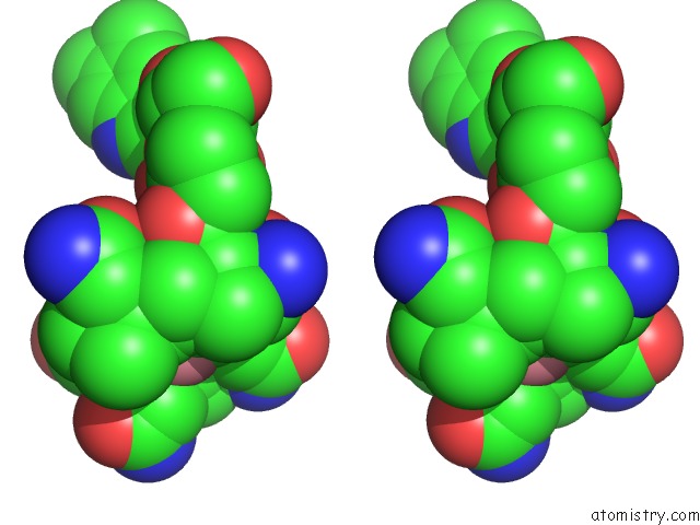
Stereo pair view
Mono view
Stereo pair view
A full contact list of Cobalt with other atoms in the Co binding
site number 1 of 1.5 Angstrom Crystal Structure of A Novel Cobalamin-Binding Protein From Desulfitobacterium Hafniense Dcb-2 within 5.0Å range:
|
Cobalt binding site 2 out of 2 in 4jgi
Go back to
Cobalt binding site 2 out
of 2 in the 1.5 Angstrom Crystal Structure of A Novel Cobalamin-Binding Protein From Desulfitobacterium Hafniense Dcb-2
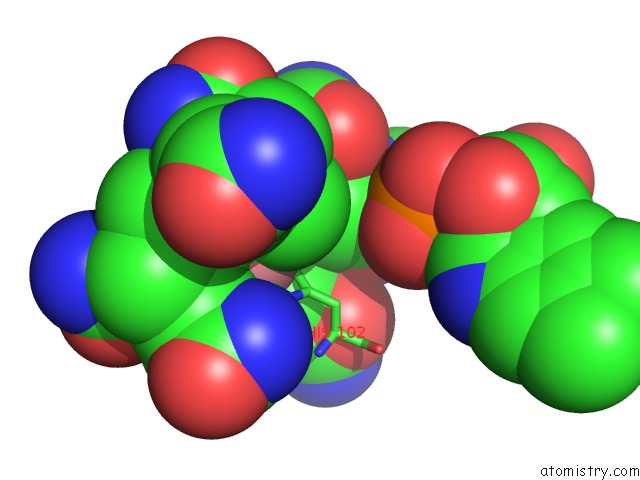
Mono view
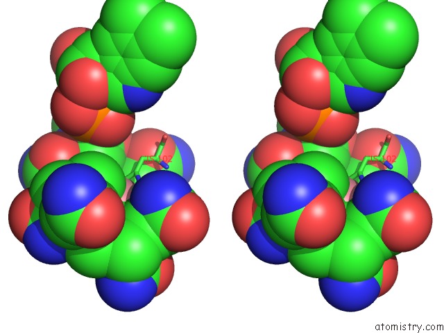
Stereo pair view
Mono view
Stereo pair view
A full contact list of Cobalt with other atoms in the Co binding
site number 2 of 1.5 Angstrom Crystal Structure of A Novel Cobalamin-Binding Protein From Desulfitobacterium Hafniense Dcb-2 within 5.0Å range:
|
Reference:
H.Sjuts,
M.S.Dunstan,
K.Fisher,
D.Leys.
Structure of the Cobalamin-Binding Protein of A Putative O-Demethylase From Desulfitobacterium Hafniense Dcb-2. Acta Crystallogr.,Sect.D V. 69 1609 2013.
ISSN: ISSN 0907-4449
PubMed: 23897483
DOI: 10.1107/S0907444913011323
Page generated: Tue Jul 30 17:14:14 2024
ISSN: ISSN 0907-4449
PubMed: 23897483
DOI: 10.1107/S0907444913011323
Last articles
Zn in 9JYWZn in 9IR4
Zn in 9IR3
Zn in 9GMX
Zn in 9GMW
Zn in 9JEJ
Zn in 9ERF
Zn in 9ERE
Zn in 9EGV
Zn in 9EGW