Cobalt »
PDB 4j2m-4ngl »
4liv »
Cobalt in PDB 4liv: Structure of Ycfd, A Ribosomal Oxygenase From Escherichia Coli in Complex with Cobalt and Succinic Acid.
Protein crystallography data
The structure of Structure of Ycfd, A Ribosomal Oxygenase From Escherichia Coli in Complex with Cobalt and Succinic Acid., PDB code: 4liv
was solved by
N.C.Brissett,
A.J.Doherty,
with X-Ray Crystallography technique. A brief refinement statistics is given in the table below:
| Resolution Low / High (Å) | 53.04 / 2.70 |
| Space group | P 43 21 2 |
| Cell size a, b, c (Å), α, β, γ (°) | 75.010, 75.010, 209.000, 90.00, 90.00, 90.00 |
| R / Rfree (%) | 19.5 / 25 |
Cobalt Binding Sites:
The binding sites of Cobalt atom in the Structure of Ycfd, A Ribosomal Oxygenase From Escherichia Coli in Complex with Cobalt and Succinic Acid.
(pdb code 4liv). This binding sites where shown within
5.0 Angstroms radius around Cobalt atom.
In total only one binding site of Cobalt was determined in the Structure of Ycfd, A Ribosomal Oxygenase From Escherichia Coli in Complex with Cobalt and Succinic Acid., PDB code: 4liv:
In total only one binding site of Cobalt was determined in the Structure of Ycfd, A Ribosomal Oxygenase From Escherichia Coli in Complex with Cobalt and Succinic Acid., PDB code: 4liv:
Cobalt binding site 1 out of 1 in 4liv
Go back to
Cobalt binding site 1 out
of 1 in the Structure of Ycfd, A Ribosomal Oxygenase From Escherichia Coli in Complex with Cobalt and Succinic Acid.
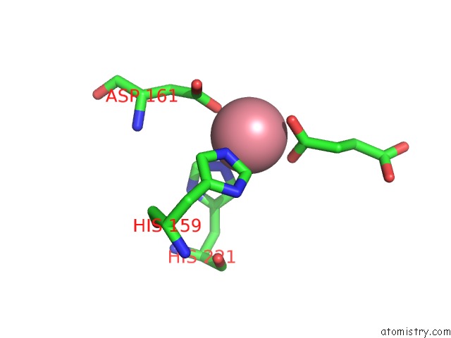
Mono view
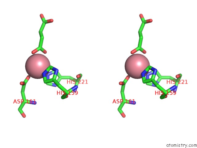
Stereo pair view
Mono view
Stereo pair view
A full contact list of Cobalt with other atoms in the Co binding
site number 1 of Structure of Ycfd, A Ribosomal Oxygenase From Escherichia Coli in Complex with Cobalt and Succinic Acid. within 5.0Å range:
|
Reference:
R.Chowdhury,
R.Sekirnik,
N.C.Brissett,
T.Krojer,
C.H.Ho,
S.S.Ng,
I.J.Clifton,
W.Ge,
N.J.Kershaw,
G.C.Fox,
J.R.Muniz,
M.Vollmar,
C.Phillips,
E.S.Pilka,
K.L.Kavanagh,
F.Von Delft,
U.Oppermann,
M.A.Mcdonough,
A.J.Doherty,
C.J.Schofield.
Ribosomal Oxygenases Are Structurally Conserved From Prokaryotes to Humans. Nature V. 509 422 2014.
ISSN: ISSN 0028-0836
PubMed: 24814345
DOI: 10.1038/NATURE13263
Page generated: Tue Jul 30 17:19:01 2024
ISSN: ISSN 0028-0836
PubMed: 24814345
DOI: 10.1038/NATURE13263
Last articles
Zn in 9JYWZn in 9IR4
Zn in 9IR3
Zn in 9GMX
Zn in 9GMW
Zn in 9JEJ
Zn in 9ERF
Zn in 9ERE
Zn in 9EGV
Zn in 9EGW