Cobalt »
PDB 4ngo-4rum »
4q3y »
Cobalt in PDB 4q3y: Crystal Structure of C. Violaceum Phenylalanine Hydroxylase D139A Mutation
Enzymatic activity of Crystal Structure of C. Violaceum Phenylalanine Hydroxylase D139A Mutation
All present enzymatic activity of Crystal Structure of C. Violaceum Phenylalanine Hydroxylase D139A Mutation:
1.14.16.1;
1.14.16.1;
Protein crystallography data
The structure of Crystal Structure of C. Violaceum Phenylalanine Hydroxylase D139A Mutation, PDB code: 4q3y
was solved by
J.A.Ronau,
M.M.Abu-Omar,
C.Das,
with X-Ray Crystallography technique. A brief refinement statistics is given in the table below:
| Resolution Low / High (Å) | 22.32 / 1.40 |
| Space group | P 1 |
| Cell size a, b, c (Å), α, β, γ (°) | 36.982, 38.672, 47.853, 76.68, 72.81, 85.54 |
| R / Rfree (%) | 16.5 / 20.5 |
Cobalt Binding Sites:
The binding sites of Cobalt atom in the Crystal Structure of C. Violaceum Phenylalanine Hydroxylase D139A Mutation
(pdb code 4q3y). This binding sites where shown within
5.0 Angstroms radius around Cobalt atom.
In total only one binding site of Cobalt was determined in the Crystal Structure of C. Violaceum Phenylalanine Hydroxylase D139A Mutation, PDB code: 4q3y:
In total only one binding site of Cobalt was determined in the Crystal Structure of C. Violaceum Phenylalanine Hydroxylase D139A Mutation, PDB code: 4q3y:
Cobalt binding site 1 out of 1 in 4q3y
Go back to
Cobalt binding site 1 out
of 1 in the Crystal Structure of C. Violaceum Phenylalanine Hydroxylase D139A Mutation
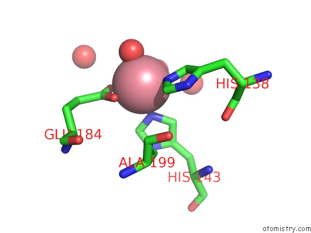
Mono view
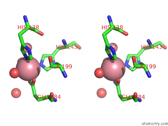
Stereo pair view
Mono view
Stereo pair view
A full contact list of Cobalt with other atoms in the Co binding
site number 1 of Crystal Structure of C. Violaceum Phenylalanine Hydroxylase D139A Mutation within 5.0Å range:
|
Reference:
J.A.Ronau,
L.N.Paul,
J.E.Fuchs,
K.R.Liedl,
M.M.Abu-Omar,
C.Das.
A Conserved Acidic Residue in Phenylalanine Hydroxylase Contributes to Cofactor Affinity and Catalysis. Biochemistry V. 53 6834 2014.
ISSN: ISSN 0006-2960
PubMed: 25295853
DOI: 10.1021/BI500734H
Page generated: Tue Jul 30 17:26:30 2024
ISSN: ISSN 0006-2960
PubMed: 25295853
DOI: 10.1021/BI500734H
Last articles
Zn in 9J0NZn in 9J0O
Zn in 9J0P
Zn in 9FJX
Zn in 9EKB
Zn in 9C0F
Zn in 9CAH
Zn in 9CH0
Zn in 9CH3
Zn in 9CH1