Cobalt »
PDB 4rut-4xc6 »
4uob »
Cobalt in PDB 4uob: Crystal Structure of Deinococcus Radiodurans Endonuclease III-3
Enzymatic activity of Crystal Structure of Deinococcus Radiodurans Endonuclease III-3
All present enzymatic activity of Crystal Structure of Deinococcus Radiodurans Endonuclease III-3:
4.2.99.18;
4.2.99.18;
Protein crystallography data
The structure of Crystal Structure of Deinococcus Radiodurans Endonuclease III-3, PDB code: 4uob
was solved by
A.Sarre,
M.Okvist,
T.Klar,
D.Hall,
A.O.Smalas,
S.Mcsweeney,
J.Timmins,
E.Moe,
with X-Ray Crystallography technique. A brief refinement statistics is given in the table below:
| Resolution Low / High (Å) | 41.932 / 1.31 |
| Space group | C 1 2 1 |
| Cell size a, b, c (Å), α, β, γ (°) | 91.240, 40.510, 72.150, 90.00, 102.34, 90.00 |
| R / Rfree (%) | 13.35 / 15.93 |
Other elements in 4uob:
The structure of Crystal Structure of Deinococcus Radiodurans Endonuclease III-3 also contains other interesting chemical elements:
| Iron | (Fe) | 4 atoms |
Cobalt Binding Sites:
The binding sites of Cobalt atom in the Crystal Structure of Deinococcus Radiodurans Endonuclease III-3
(pdb code 4uob). This binding sites where shown within
5.0 Angstroms radius around Cobalt atom.
In total only one binding site of Cobalt was determined in the Crystal Structure of Deinococcus Radiodurans Endonuclease III-3, PDB code: 4uob:
In total only one binding site of Cobalt was determined in the Crystal Structure of Deinococcus Radiodurans Endonuclease III-3, PDB code: 4uob:
Cobalt binding site 1 out of 1 in 4uob
Go back to
Cobalt binding site 1 out
of 1 in the Crystal Structure of Deinococcus Radiodurans Endonuclease III-3
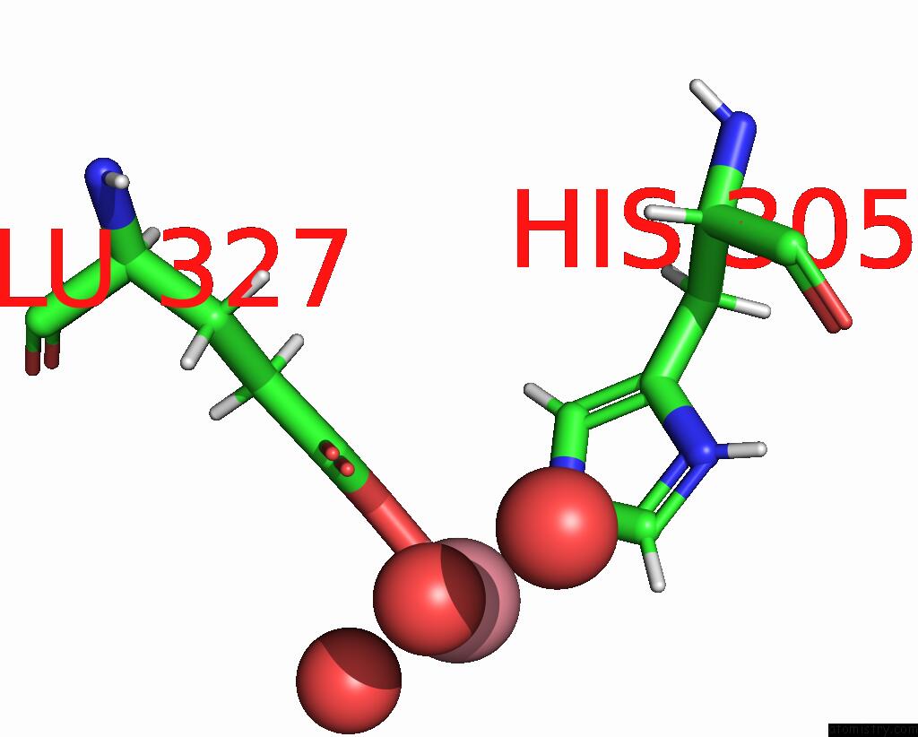
Mono view
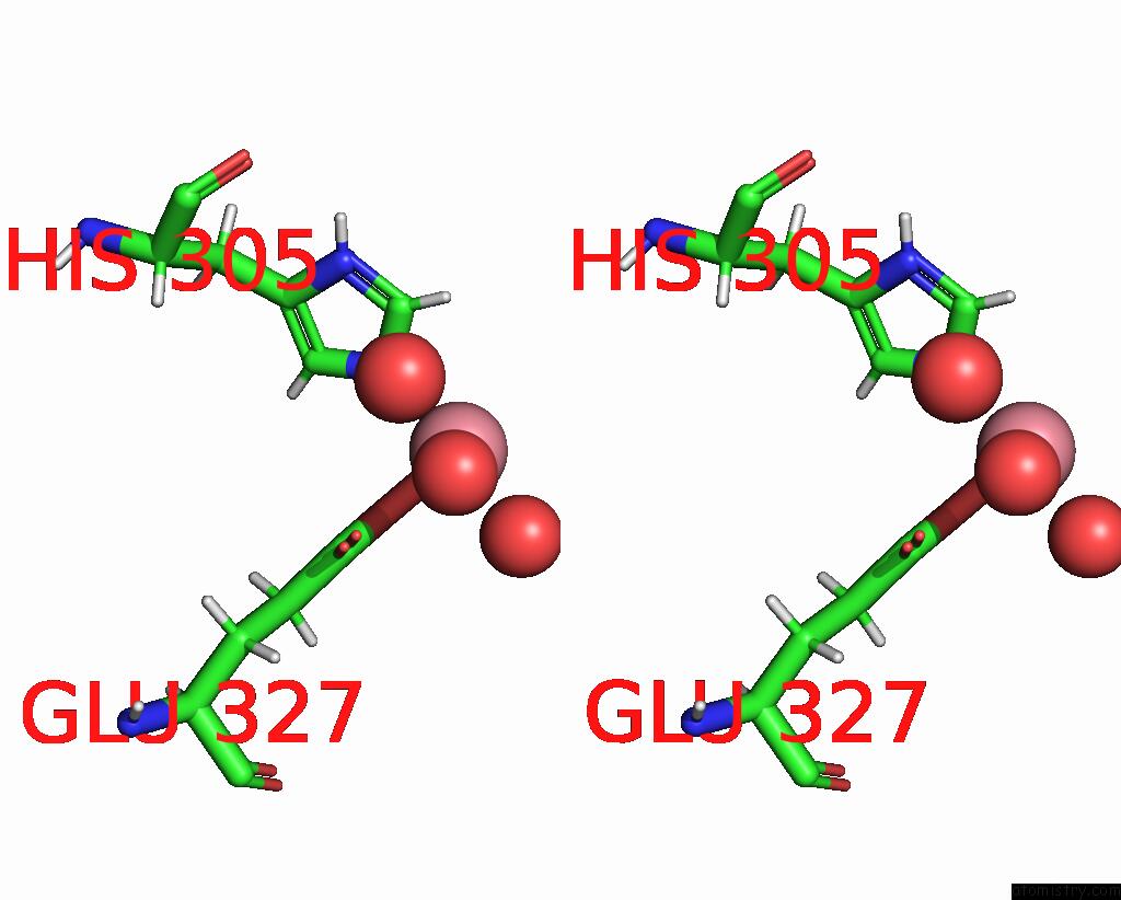
Stereo pair view
Mono view
Stereo pair view
A full contact list of Cobalt with other atoms in the Co binding
site number 1 of Crystal Structure of Deinococcus Radiodurans Endonuclease III-3 within 5.0Å range:
|
Reference:
A.Sarre,
M.Okvist,
T.Klar,
D.R.Hall,
A.O.Smalas,
S.Mcsweeney,
J.Timmins,
E.Moe.
Structural and Functional Characterization of Two Unusual Endonuclease III Enzymes From Deinococcus Radiodurans. J.Struct.Biol. V. 191 87 2015.
ISSN: ISSN 1047-8477
PubMed: 26172070
DOI: 10.1016/J.JSB.2015.05.009
Page generated: Tue Jul 30 17:33:44 2024
ISSN: ISSN 1047-8477
PubMed: 26172070
DOI: 10.1016/J.JSB.2015.05.009
Last articles
Zn in 9MJ5Zn in 9HNW
Zn in 9G0L
Zn in 9FNE
Zn in 9DZN
Zn in 9E0I
Zn in 9D32
Zn in 9DAK
Zn in 8ZXC
Zn in 8ZUF