Cobalt »
PDB 4xim-5d6e »
5azr »
Cobalt in PDB 5azr: Crystal Structure of Aqua-Cobalt(III) Tetradehydrocorrin in the Heme Pocket of Horse Heart Myoglobin
Protein crystallography data
The structure of Crystal Structure of Aqua-Cobalt(III) Tetradehydrocorrin in the Heme Pocket of Horse Heart Myoglobin, PDB code: 5azr
was solved by
E.Mizohata,
Y.Morita,
K.Oohora,
T.Inoue,
T.Hayashi,
with X-Ray Crystallography technique. A brief refinement statistics is given in the table below:
| Resolution Low / High (Å) | 50.00 / 1.20 |
| Space group | P 1 21 1 |
| Cell size a, b, c (Å), α, β, γ (°) | 34.786, 28.837, 63.324, 90.00, 106.17, 90.00 |
| R / Rfree (%) | 12.1 / 16.8 |
Cobalt Binding Sites:
The binding sites of Cobalt atom in the Crystal Structure of Aqua-Cobalt(III) Tetradehydrocorrin in the Heme Pocket of Horse Heart Myoglobin
(pdb code 5azr). This binding sites where shown within
5.0 Angstroms radius around Cobalt atom.
In total 2 binding sites of Cobalt where determined in the Crystal Structure of Aqua-Cobalt(III) Tetradehydrocorrin in the Heme Pocket of Horse Heart Myoglobin, PDB code: 5azr:
Jump to Cobalt binding site number: 1; 2;
In total 2 binding sites of Cobalt where determined in the Crystal Structure of Aqua-Cobalt(III) Tetradehydrocorrin in the Heme Pocket of Horse Heart Myoglobin, PDB code: 5azr:
Jump to Cobalt binding site number: 1; 2;
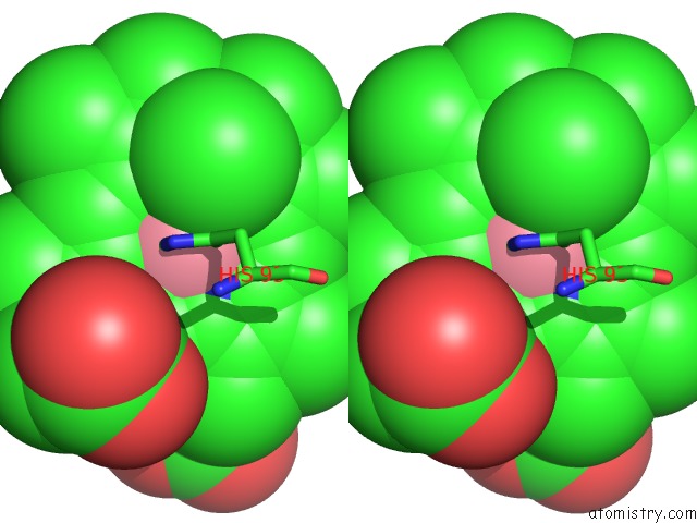
Cobalt binding site 1 out of 2 in 5azr
Go back to
Cobalt binding site 1 out
of 2 in the Crystal Structure of Aqua-Cobalt(III) Tetradehydrocorrin in the Heme Pocket of Horse Heart Myoglobin
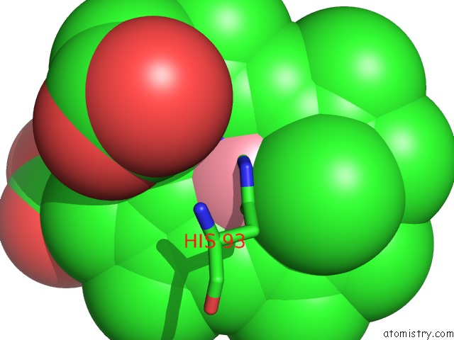
Mono view

Stereo pair view
Mono view
Stereo pair view
A full contact list of Cobalt with other atoms in the Co binding
site number 1 of Crystal Structure of Aqua-Cobalt(III) Tetradehydrocorrin in the Heme Pocket of Horse Heart Myoglobin within 5.0Å range:
|
Cobalt binding site 2 out of 2 in 5azr
Go back to
Cobalt binding site 2 out
of 2 in the Crystal Structure of Aqua-Cobalt(III) Tetradehydrocorrin in the Heme Pocket of Horse Heart Myoglobin

Mono view
Stereo pair view
Mono view
Stereo pair view
A full contact list of Cobalt with other atoms in the Co binding
site number 2 of Crystal Structure of Aqua-Cobalt(III) Tetradehydrocorrin in the Heme Pocket of Horse Heart Myoglobin within 5.0Å range:
|
Reference:
Y.Morita,
K.Oohora,
E.Mizohata,
A.Sawada,
T.Kamachi,
K.Yoshizawa,
T.Inoue,
T.Hayashi.
Crystal Structures and Coordination Behavior of Aqua- and Cyano-Co(III) Tetradehydrocorrins in the Heme Pocket of Myoglobin Inorg.Chem. V. 55 1287 2016.
ISSN: ISSN 0020-1669
PubMed: 26760442
DOI: 10.1021/ACS.INORGCHEM.5B02598
Page generated: Tue Jul 30 17:43:14 2024
ISSN: ISSN 0020-1669
PubMed: 26760442
DOI: 10.1021/ACS.INORGCHEM.5B02598
Last articles
Zn in 9MJ5Zn in 9HNW
Zn in 9G0L
Zn in 9FNE
Zn in 9DZN
Zn in 9E0I
Zn in 9D32
Zn in 9DAK
Zn in 8ZXC
Zn in 8ZUF