Cobalt »
PDB 5d6f-5ikv »
5e8g »
Cobalt in PDB 5e8g: Crystal Structure of the Dna Binding Domain of Human Transcription Factor FLI1
Protein crystallography data
The structure of Crystal Structure of the Dna Binding Domain of Human Transcription Factor FLI1, PDB code: 5e8g
was solved by
C.Hou,
O.V.Tsodikov,
with X-Ray Crystallography technique. A brief refinement statistics is given in the table below:
| Resolution Low / High (Å) | 40.00 / 2.70 |
| Space group | H 3 |
| Cell size a, b, c (Å), α, β, γ (°) | 140.551, 140.551, 85.140, 90.00, 90.00, 120.00 |
| R / Rfree (%) | 19.6 / 24.8 |
Cobalt Binding Sites:
The binding sites of Cobalt atom in the Crystal Structure of the Dna Binding Domain of Human Transcription Factor FLI1
(pdb code 5e8g). This binding sites where shown within
5.0 Angstroms radius around Cobalt atom.
In total 4 binding sites of Cobalt where determined in the Crystal Structure of the Dna Binding Domain of Human Transcription Factor FLI1, PDB code: 5e8g:
Jump to Cobalt binding site number: 1; 2; 3; 4;
In total 4 binding sites of Cobalt where determined in the Crystal Structure of the Dna Binding Domain of Human Transcription Factor FLI1, PDB code: 5e8g:
Jump to Cobalt binding site number: 1; 2; 3; 4;
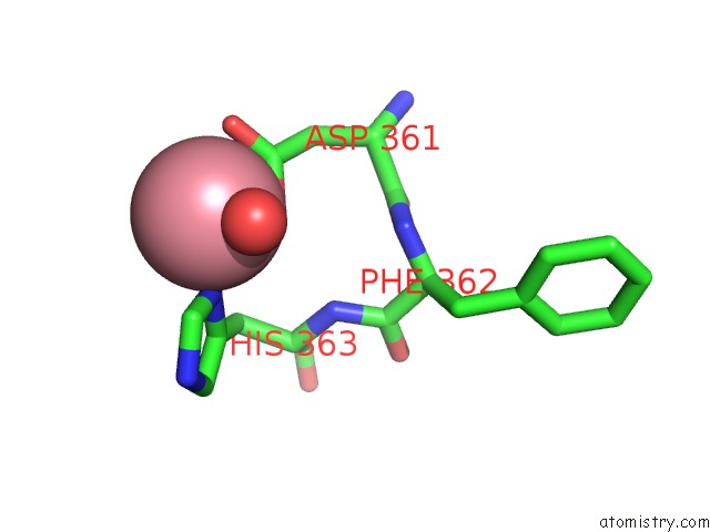
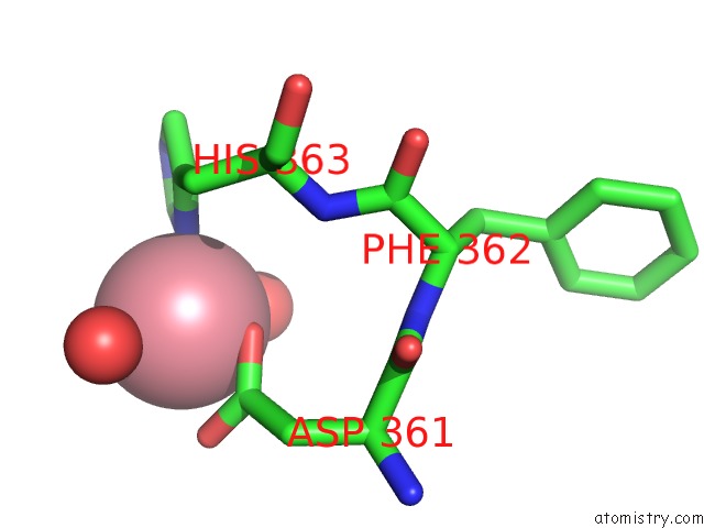
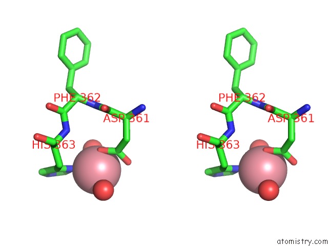
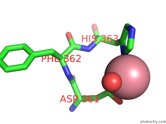
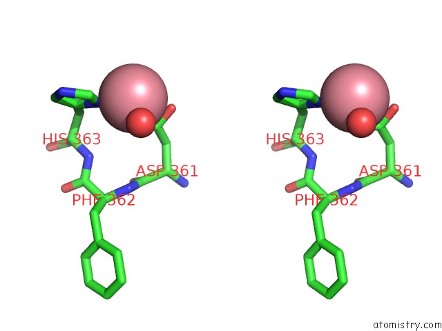
Cobalt binding site 1 out of 4 in 5e8g
Go back to
Cobalt binding site 1 out
of 4 in the Crystal Structure of the Dna Binding Domain of Human Transcription Factor FLI1

Mono view

Stereo pair view
Mono view
Stereo pair view
A full contact list of Cobalt with other atoms in the Co binding
site number 1 of Crystal Structure of the Dna Binding Domain of Human Transcription Factor FLI1 within 5.0Å range:
|
Cobalt binding site 2 out of 4 in 5e8g
Go back to
Cobalt binding site 2 out
of 4 in the Crystal Structure of the Dna Binding Domain of Human Transcription Factor FLI1
Mono view

Stereo pair view
Mono view
Stereo pair view
A full contact list of Cobalt with other atoms in the Co binding
site number 2 of Crystal Structure of the Dna Binding Domain of Human Transcription Factor FLI1 within 5.0Å range:
|
Cobalt binding site 3 out of 4 in 5e8g
Go back to
Cobalt binding site 3 out
of 4 in the Crystal Structure of the Dna Binding Domain of Human Transcription Factor FLI1
Mono view
Stereo pair view
Mono view
Stereo pair view
A full contact list of Cobalt with other atoms in the Co binding
site number 3 of Crystal Structure of the Dna Binding Domain of Human Transcription Factor FLI1 within 5.0Å range:
|
Cobalt binding site 4 out of 4 in 5e8g
Go back to
Cobalt binding site 4 out
of 4 in the Crystal Structure of the Dna Binding Domain of Human Transcription Factor FLI1
Mono view
Stereo pair view
Mono view
Stereo pair view
A full contact list of Cobalt with other atoms in the Co binding
site number 4 of Crystal Structure of the Dna Binding Domain of Human Transcription Factor FLI1 within 5.0Å range:
|
Reference:
C.Hou,
O.V.Tsodikov.
Structural Basis For Dimerization and Dna Binding of Transcription Factor FLI1. Biochemistry V. 54 7365 2015.
ISSN: ISSN 0006-2960
PubMed: 26618620
DOI: 10.1021/ACS.BIOCHEM.5B01121
Page generated: Sun Jul 13 20:19:58 2025
ISSN: ISSN 0006-2960
PubMed: 26618620
DOI: 10.1021/ACS.BIOCHEM.5B01121
Last articles
Cu in 6RHUCu in 6RHR
Cu in 6RHP
Cu in 6RHI
Cu in 6RHO
Cu in 6RHH
Cu in 6QYB
Cu in 6Q6B
Cu in 6RGP
Cu in 6RGH