Cobalt »
PDB 5d6f-5ikv »
5f1a »
Cobalt in PDB 5f1a: The Crystal Structure of Salicylate Bound to Human Cyclooxygenase-2
Enzymatic activity of The Crystal Structure of Salicylate Bound to Human Cyclooxygenase-2
All present enzymatic activity of The Crystal Structure of Salicylate Bound to Human Cyclooxygenase-2:
1.14.99.1;
1.14.99.1;
Protein crystallography data
The structure of The Crystal Structure of Salicylate Bound to Human Cyclooxygenase-2, PDB code: 5f1a
was solved by
M.J.Lucido,
B.J.Orlando,
M.G.Malkowski,
with X-Ray Crystallography technique. A brief refinement statistics is given in the table below:
| Resolution Low / High (Å) | 33.32 / 2.38 |
| Space group | I 2 2 2 |
| Cell size a, b, c (Å), α, β, γ (°) | 118.410, 132.660, 178.740, 90.00, 90.00, 90.00 |
| R / Rfree (%) | 17.4 / 21.8 |
Cobalt Binding Sites:
The binding sites of Cobalt atom in the The Crystal Structure of Salicylate Bound to Human Cyclooxygenase-2
(pdb code 5f1a). This binding sites where shown within
5.0 Angstroms radius around Cobalt atom.
In total 2 binding sites of Cobalt where determined in the The Crystal Structure of Salicylate Bound to Human Cyclooxygenase-2, PDB code: 5f1a:
Jump to Cobalt binding site number: 1; 2;
In total 2 binding sites of Cobalt where determined in the The Crystal Structure of Salicylate Bound to Human Cyclooxygenase-2, PDB code: 5f1a:
Jump to Cobalt binding site number: 1; 2;
Cobalt binding site 1 out of 2 in 5f1a
Go back to
Cobalt binding site 1 out
of 2 in the The Crystal Structure of Salicylate Bound to Human Cyclooxygenase-2
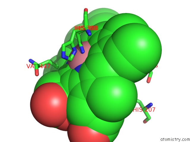
Mono view
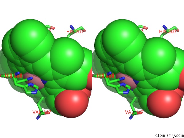
Stereo pair view
Mono view
Stereo pair view
A full contact list of Cobalt with other atoms in the Co binding
site number 1 of The Crystal Structure of Salicylate Bound to Human Cyclooxygenase-2 within 5.0Å range:
|
Cobalt binding site 2 out of 2 in 5f1a
Go back to
Cobalt binding site 2 out
of 2 in the The Crystal Structure of Salicylate Bound to Human Cyclooxygenase-2
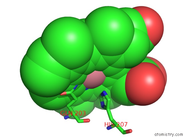
Mono view
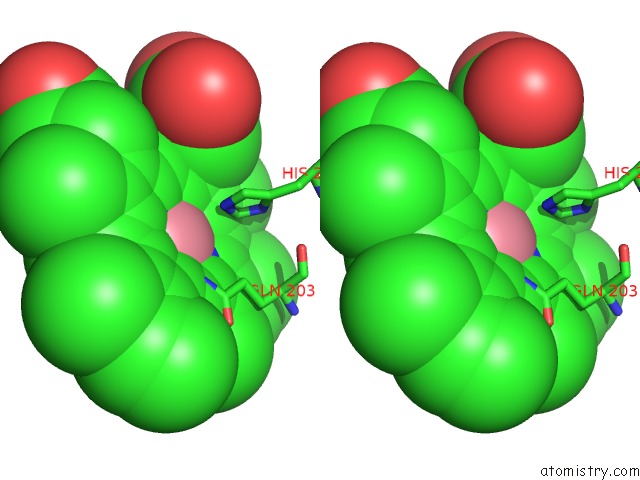
Stereo pair view
Mono view
Stereo pair view
A full contact list of Cobalt with other atoms in the Co binding
site number 2 of The Crystal Structure of Salicylate Bound to Human Cyclooxygenase-2 within 5.0Å range:
|
Reference:
M.J.Lucido,
B.J.Orlando,
A.J.Vecchio,
M.G.Malkowski.
Crystal Structure of Aspirin-Acetylated Human Cyclooxygenase-2: Insight Into the Formation of Products with Reversed Stereochemistry. Biochemistry V. 55 1226 2016.
ISSN: ISSN 0006-2960
PubMed: 26859324
DOI: 10.1021/ACS.BIOCHEM.5B01378
Page generated: Sun Jul 13 20:22:10 2025
ISSN: ISSN 0006-2960
PubMed: 26859324
DOI: 10.1021/ACS.BIOCHEM.5B01378
Last articles
Cu in 5ZP2Cu in 5ZP1
Cu in 5ZP0
Cu in 5ZOZ
Cu in 5ZOY
Cu in 5ZL1
Cu in 5ZOX
Cu in 5ZOW
Cu in 5ZLM
Cu in 5ZOU