Cobalt »
PDB 5img-5np4 »
5kir »
Cobalt in PDB 5kir: The Structure of Vioxx Bound to Human Cox-2
Enzymatic activity of The Structure of Vioxx Bound to Human Cox-2
All present enzymatic activity of The Structure of Vioxx Bound to Human Cox-2:
1.14.99.1;
1.14.99.1;
Protein crystallography data
The structure of The Structure of Vioxx Bound to Human Cox-2, PDB code: 5kir
was solved by
B.J.Orlando,
M.G.Malkowski,
with X-Ray Crystallography technique. A brief refinement statistics is given in the table below:
| Resolution Low / High (Å) | 30.03 / 2.70 |
| Space group | I 2 2 2 |
| Cell size a, b, c (Å), α, β, γ (°) | 126.989, 149.422, 185.055, 90.00, 90.00, 90.00 |
| R / Rfree (%) | 17.8 / 22 |
Cobalt Binding Sites:
The binding sites of Cobalt atom in the The Structure of Vioxx Bound to Human Cox-2
(pdb code 5kir). This binding sites where shown within
5.0 Angstroms radius around Cobalt atom.
In total 2 binding sites of Cobalt where determined in the The Structure of Vioxx Bound to Human Cox-2, PDB code: 5kir:
Jump to Cobalt binding site number: 1; 2;
In total 2 binding sites of Cobalt where determined in the The Structure of Vioxx Bound to Human Cox-2, PDB code: 5kir:
Jump to Cobalt binding site number: 1; 2;
Cobalt binding site 1 out of 2 in 5kir
Go back to
Cobalt binding site 1 out
of 2 in the The Structure of Vioxx Bound to Human Cox-2
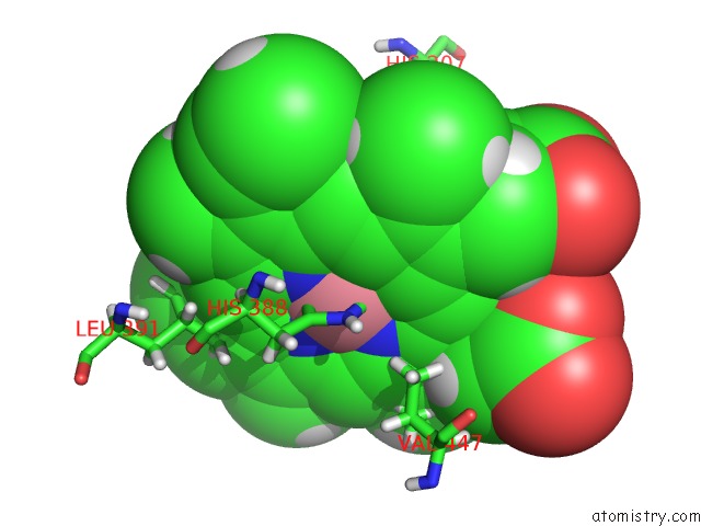
Mono view
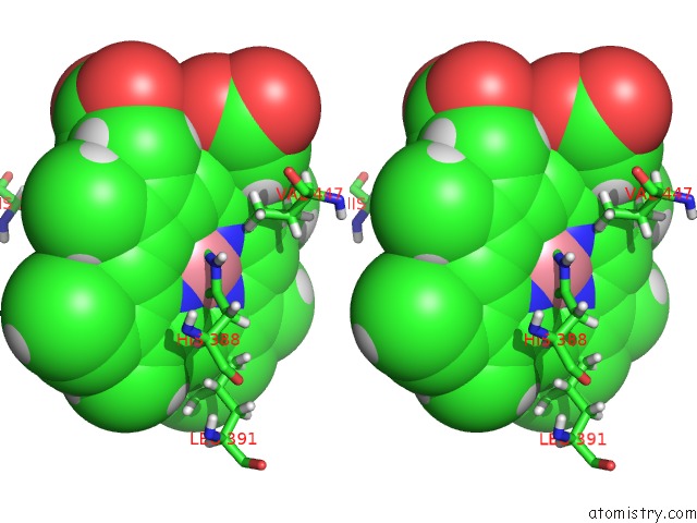
Stereo pair view
Mono view
Stereo pair view
A full contact list of Cobalt with other atoms in the Co binding
site number 1 of The Structure of Vioxx Bound to Human Cox-2 within 5.0Å range:
|
Cobalt binding site 2 out of 2 in 5kir
Go back to
Cobalt binding site 2 out
of 2 in the The Structure of Vioxx Bound to Human Cox-2
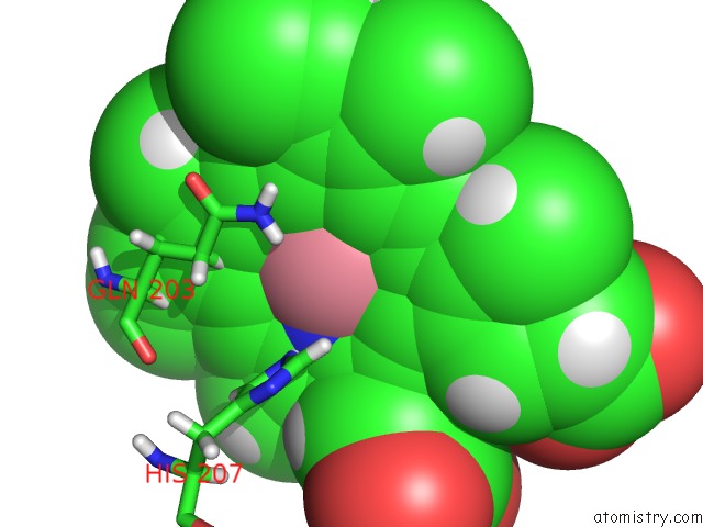
Mono view
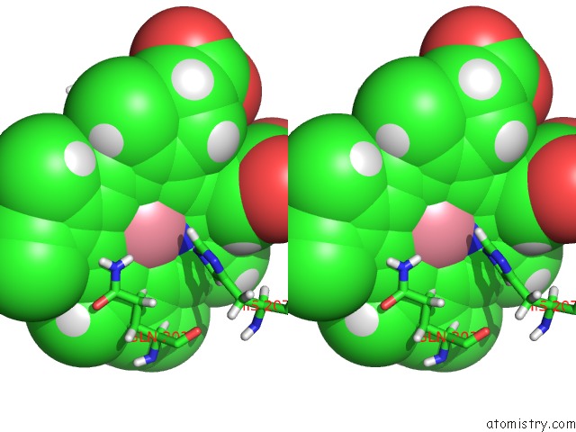
Stereo pair view
Mono view
Stereo pair view
A full contact list of Cobalt with other atoms in the Co binding
site number 2 of The Structure of Vioxx Bound to Human Cox-2 within 5.0Å range:
|
Reference:
B.J.Orlando,
M.G.Malkowski.
Crystal Structure of Rofecoxib Bound to Human Cyclooxygenase-2. Acta Crystallogr.,Sect.F V. 72 772 2016.
ISSN: ESSN 2053-230X
PubMed: 27710942
DOI: 10.1107/S2053230X16014230
Page generated: Tue Jul 30 18:01:41 2024
ISSN: ESSN 2053-230X
PubMed: 27710942
DOI: 10.1107/S2053230X16014230
Last articles
Zn in 9J0NZn in 9J0O
Zn in 9J0P
Zn in 9FJX
Zn in 9EKB
Zn in 9C0F
Zn in 9CAH
Zn in 9CH0
Zn in 9CH3
Zn in 9CH1