Cobalt »
PDB 5img-5np4 »
5m3b »
Cobalt in PDB 5m3b: Structure of Cobinamide-Bound Btuf Mutant W66L, the Periplasmic Vitamin B12 Binding Protein in E.Coli
Protein crystallography data
The structure of Structure of Cobinamide-Bound Btuf Mutant W66L, the Periplasmic Vitamin B12 Binding Protein in E.Coli, PDB code: 5m3b
was solved by
S.A.Mireku,
M.Ruetz,
T.Zhou,
V.M.Korkhov,
B.Kraeutler,
K.P.Locher,
with X-Ray Crystallography technique. A brief refinement statistics is given in the table below:
| Resolution Low / High (Å) | 19.67 / 1.50 |
| Space group | C 1 2 1 |
| Cell size a, b, c (Å), α, β, γ (°) | 136.280, 90.460, 50.810, 90.00, 110.74, 90.00 |
| R / Rfree (%) | 19.6 / 22.9 |
Cobalt Binding Sites:
The binding sites of Cobalt atom in the Structure of Cobinamide-Bound Btuf Mutant W66L, the Periplasmic Vitamin B12 Binding Protein in E.Coli
(pdb code 5m3b). This binding sites where shown within
5.0 Angstroms radius around Cobalt atom.
In total 2 binding sites of Cobalt where determined in the Structure of Cobinamide-Bound Btuf Mutant W66L, the Periplasmic Vitamin B12 Binding Protein in E.Coli, PDB code: 5m3b:
Jump to Cobalt binding site number: 1; 2;
In total 2 binding sites of Cobalt where determined in the Structure of Cobinamide-Bound Btuf Mutant W66L, the Periplasmic Vitamin B12 Binding Protein in E.Coli, PDB code: 5m3b:
Jump to Cobalt binding site number: 1; 2;
Cobalt binding site 1 out of 2 in 5m3b
Go back to
Cobalt binding site 1 out
of 2 in the Structure of Cobinamide-Bound Btuf Mutant W66L, the Periplasmic Vitamin B12 Binding Protein in E.Coli
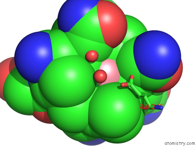
Mono view
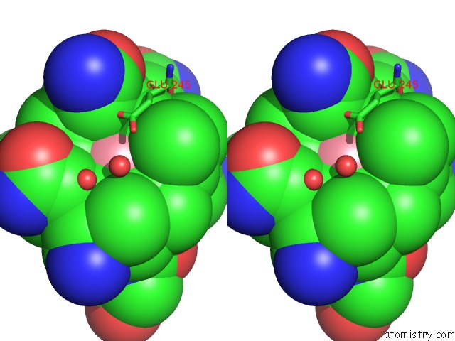
Stereo pair view
Mono view
Stereo pair view
A full contact list of Cobalt with other atoms in the Co binding
site number 1 of Structure of Cobinamide-Bound Btuf Mutant W66L, the Periplasmic Vitamin B12 Binding Protein in E.Coli within 5.0Å range:
|
Cobalt binding site 2 out of 2 in 5m3b
Go back to
Cobalt binding site 2 out
of 2 in the Structure of Cobinamide-Bound Btuf Mutant W66L, the Periplasmic Vitamin B12 Binding Protein in E.Coli
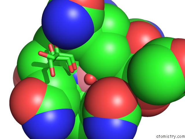
Mono view
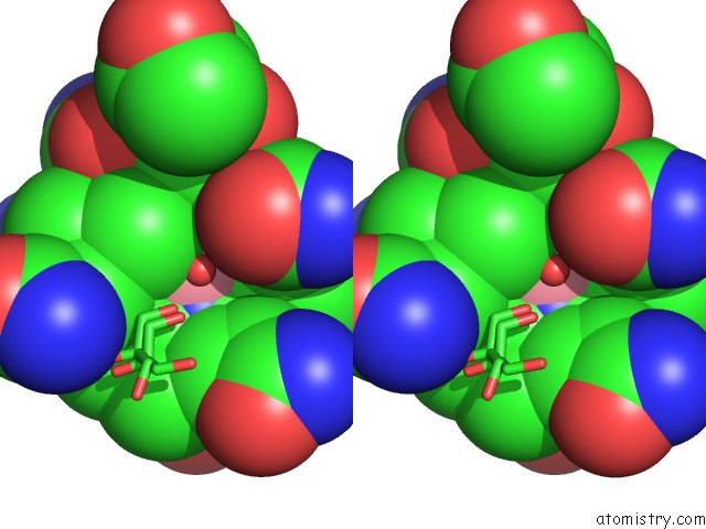
Stereo pair view
Mono view
Stereo pair view
A full contact list of Cobalt with other atoms in the Co binding
site number 2 of Structure of Cobinamide-Bound Btuf Mutant W66L, the Periplasmic Vitamin B12 Binding Protein in E.Coli within 5.0Å range:
|
Reference:
S.A.Mireku,
M.Ruetz,
T.Zhou,
V.M.Korkhov,
B.Krautler,
K.P.Locher.
Conformational Change of A Tryptophan Residue in Btuf Facilitates Binding and Transport of Cobinamide By the Vitamin B12 Transporter Btucd-F. Sci Rep V. 7 41575 2017.
ISSN: ESSN 2045-2322
PubMed: 28128319
DOI: 10.1038/SREP41575
Page generated: Tue Jul 30 18:05:23 2024
ISSN: ESSN 2045-2322
PubMed: 28128319
DOI: 10.1038/SREP41575
Last articles
Zn in 9J0NZn in 9J0O
Zn in 9J0P
Zn in 9FJX
Zn in 9EKB
Zn in 9C0F
Zn in 9CAH
Zn in 9CH0
Zn in 9CH3
Zn in 9CH1