Cobalt »
PDB 5vtd-5zt2 »
5yka »
Cobalt in PDB 5yka: Crystal Structure of the Kdo Hydroxylase Kdoo, A Non-Heme Fe(II) Alphaketoglutarate Dependent Dioxygenase in Complex with Cobalt(II)
Protein crystallography data
The structure of Crystal Structure of the Kdo Hydroxylase Kdoo, A Non-Heme Fe(II) Alphaketoglutarate Dependent Dioxygenase in Complex with Cobalt(II), PDB code: 5yka
was solved by
H.S.Chung,
C.W.Pemble,
S.H.Joo,
C.R.Raetz,
with X-Ray Crystallography technique. A brief refinement statistics is given in the table below:
| Resolution Low / High (Å) | 23.64 / 1.45 |
| Space group | P 21 21 21 |
| Cell size a, b, c (Å), α, β, γ (°) | 45.802, 59.607, 116.438, 90.00, 90.00, 90.00 |
| R / Rfree (%) | 15.7 / 17.9 |
Other elements in 5yka:
The structure of Crystal Structure of the Kdo Hydroxylase Kdoo, A Non-Heme Fe(II) Alphaketoglutarate Dependent Dioxygenase in Complex with Cobalt(II) also contains other interesting chemical elements:
| Chlorine | (Cl) | 2 atoms |
Cobalt Binding Sites:
The binding sites of Cobalt atom in the Crystal Structure of the Kdo Hydroxylase Kdoo, A Non-Heme Fe(II) Alphaketoglutarate Dependent Dioxygenase in Complex with Cobalt(II)
(pdb code 5yka). This binding sites where shown within
5.0 Angstroms radius around Cobalt atom.
In total only one binding site of Cobalt was determined in the Crystal Structure of the Kdo Hydroxylase Kdoo, A Non-Heme Fe(II) Alphaketoglutarate Dependent Dioxygenase in Complex with Cobalt(II), PDB code: 5yka:
In total only one binding site of Cobalt was determined in the Crystal Structure of the Kdo Hydroxylase Kdoo, A Non-Heme Fe(II) Alphaketoglutarate Dependent Dioxygenase in Complex with Cobalt(II), PDB code: 5yka:
Cobalt binding site 1 out of 1 in 5yka
Go back to
Cobalt binding site 1 out
of 1 in the Crystal Structure of the Kdo Hydroxylase Kdoo, A Non-Heme Fe(II) Alphaketoglutarate Dependent Dioxygenase in Complex with Cobalt(II)
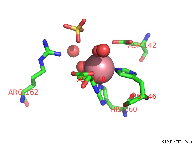
Mono view
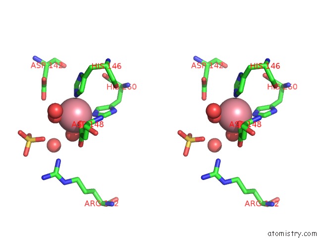
Stereo pair view
Mono view
Stereo pair view
A full contact list of Cobalt with other atoms in the Co binding
site number 1 of Crystal Structure of the Kdo Hydroxylase Kdoo, A Non-Heme Fe(II) Alphaketoglutarate Dependent Dioxygenase in Complex with Cobalt(II) within 5.0Å range:
|
Reference:
S.H.Joo,
C.W.Pemble,
E.G.Yang,
C.R.H.Raetz,
H.S.Chung.
Biochemical and Structural Insights Into An Fe(II)/ Alpha-Ketoglutarate/O2-Dependent Dioxygenase, Kdo 3-Hydroxylase (Kdoo). J. Mol. Biol. V. 430 4036 2018.
ISSN: ESSN 1089-8638
PubMed: 30092253
DOI: 10.1016/J.JMB.2018.07.029
Page generated: Tue Jul 30 18:19:28 2024
ISSN: ESSN 1089-8638
PubMed: 30092253
DOI: 10.1016/J.JMB.2018.07.029
Last articles
Zn in 9J0NZn in 9J0O
Zn in 9J0P
Zn in 9FJX
Zn in 9EKB
Zn in 9C0F
Zn in 9CAH
Zn in 9CH0
Zn in 9CH3
Zn in 9CH1