Cobalt »
PDB 5zt7-6eg9 »
6bnx »
Cobalt in PDB 6bnx: Crystal Structure of V278E-Glyoxalase I Mutant From Zea Mays in Space Group P6(3)
Enzymatic activity of Crystal Structure of V278E-Glyoxalase I Mutant From Zea Mays in Space Group P6(3)
All present enzymatic activity of Crystal Structure of V278E-Glyoxalase I Mutant From Zea Mays in Space Group P6(3):
4.4.1.5;
4.4.1.5;
Protein crystallography data
The structure of Crystal Structure of V278E-Glyoxalase I Mutant From Zea Mays in Space Group P6(3), PDB code: 6bnx
was solved by
C.E.Alvarez,
R.B.Agostini,
J.M.Gonzalez,
M.F.Drincovich,
V.A.Camposbermudez,
S.Klinke,
with X-Ray Crystallography technique. A brief refinement statistics is given in the table below:
| Resolution Low / High (Å) | 41.05 / 1.80 |
| Space group | P 63 |
| Cell size a, b, c (Å), α, β, γ (°) | 82.110, 82.110, 74.970, 90.00, 90.00, 120.00 |
| R / Rfree (%) | 18.3 / 22.4 |
Cobalt Binding Sites:
The binding sites of Cobalt atom in the Crystal Structure of V278E-Glyoxalase I Mutant From Zea Mays in Space Group P6(3)
(pdb code 6bnx). This binding sites where shown within
5.0 Angstroms radius around Cobalt atom.
In total only one binding site of Cobalt was determined in the Crystal Structure of V278E-Glyoxalase I Mutant From Zea Mays in Space Group P6(3), PDB code: 6bnx:
In total only one binding site of Cobalt was determined in the Crystal Structure of V278E-Glyoxalase I Mutant From Zea Mays in Space Group P6(3), PDB code: 6bnx:
Cobalt binding site 1 out of 1 in 6bnx
Go back to
Cobalt binding site 1 out
of 1 in the Crystal Structure of V278E-Glyoxalase I Mutant From Zea Mays in Space Group P6(3)
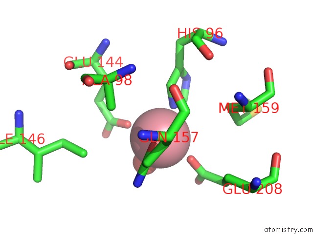
Mono view
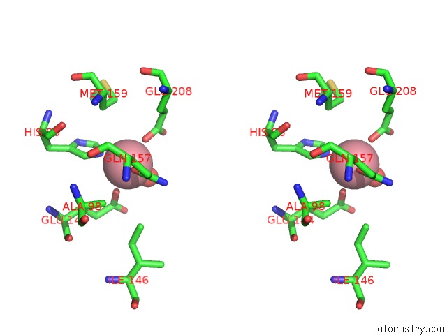
Stereo pair view
Mono view
Stereo pair view
A full contact list of Cobalt with other atoms in the Co binding
site number 1 of Crystal Structure of V278E-Glyoxalase I Mutant From Zea Mays in Space Group P6(3) within 5.0Å range:
|
Reference:
J.M.Gonzalez,
R.B.Agostini,
C.E.Alvarez,
S.Klinke,
C.S.Andreo,
V.A.Campos-Bermudez.
Deciphering the Number and Location of Active Sites in the Monomeric Glyoxalase I of Zea Mays. Febs J. V. 286 3255 2019.
ISSN: ISSN 1742-464X
PubMed: 30993890
DOI: 10.1111/FEBS.14855
Page generated: Tue Jul 30 18:28:55 2024
ISSN: ISSN 1742-464X
PubMed: 30993890
DOI: 10.1111/FEBS.14855
Last articles
Zn in 9J0NZn in 9J0O
Zn in 9J0P
Zn in 9FJX
Zn in 9EKB
Zn in 9C0F
Zn in 9CAH
Zn in 9CH0
Zn in 9CH3
Zn in 9CH1