Cobalt »
PDB 6kgh-6oxc »
6nnc »
Cobalt in PDB 6nnc: Structure of Dihydrofolate Reductase From Mycobacterium Tuberculosis in Complex with Nadph and Pemetrexed
Enzymatic activity of Structure of Dihydrofolate Reductase From Mycobacterium Tuberculosis in Complex with Nadph and Pemetrexed
All present enzymatic activity of Structure of Dihydrofolate Reductase From Mycobacterium Tuberculosis in Complex with Nadph and Pemetrexed:
1.5.1.3;
1.5.1.3;
Protein crystallography data
The structure of Structure of Dihydrofolate Reductase From Mycobacterium Tuberculosis in Complex with Nadph and Pemetrexed, PDB code: 6nnc
was solved by
J.A.Ribeiro,
S.M.Chavez-Pacheco,
M.V.B.Dias,
with X-Ray Crystallography technique. A brief refinement statistics is given in the table below:
| Resolution Low / High (Å) | 32.16 / 1.80 |
| Space group | P 21 21 21 |
| Cell size a, b, c (Å), α, β, γ (°) | 61.346, 70.718, 72.216, 90.00, 90.00, 90.00 |
| R / Rfree (%) | 16.1 / 20.4 |
Cobalt Binding Sites:
The binding sites of Cobalt atom in the Structure of Dihydrofolate Reductase From Mycobacterium Tuberculosis in Complex with Nadph and Pemetrexed
(pdb code 6nnc). This binding sites where shown within
5.0 Angstroms radius around Cobalt atom.
In total 2 binding sites of Cobalt where determined in the Structure of Dihydrofolate Reductase From Mycobacterium Tuberculosis in Complex with Nadph and Pemetrexed, PDB code: 6nnc:
Jump to Cobalt binding site number: 1; 2;
In total 2 binding sites of Cobalt where determined in the Structure of Dihydrofolate Reductase From Mycobacterium Tuberculosis in Complex with Nadph and Pemetrexed, PDB code: 6nnc:
Jump to Cobalt binding site number: 1; 2;
Cobalt binding site 1 out of 2 in 6nnc
Go back to
Cobalt binding site 1 out
of 2 in the Structure of Dihydrofolate Reductase From Mycobacterium Tuberculosis in Complex with Nadph and Pemetrexed
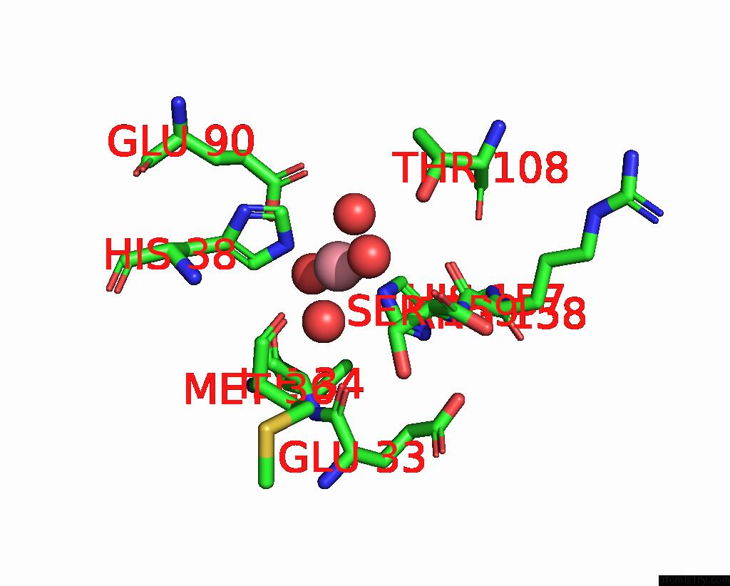
Mono view
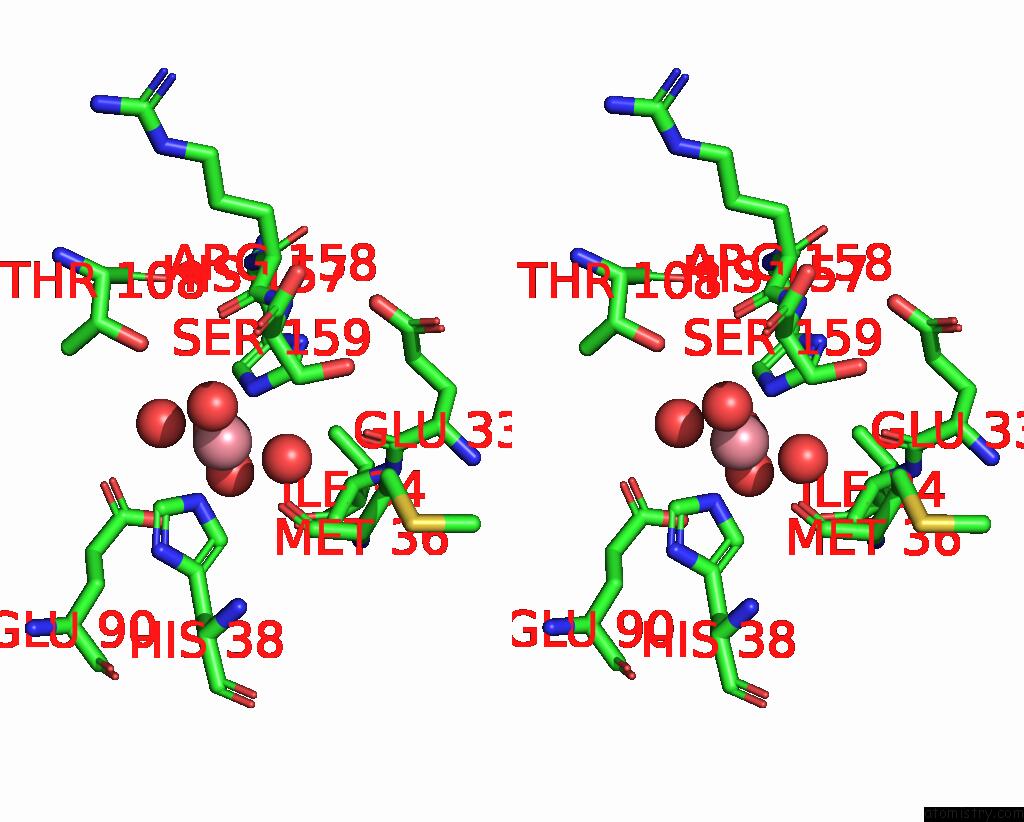
Stereo pair view
Mono view
Stereo pair view
A full contact list of Cobalt with other atoms in the Co binding
site number 1 of Structure of Dihydrofolate Reductase From Mycobacterium Tuberculosis in Complex with Nadph and Pemetrexed within 5.0Å range:
|
Cobalt binding site 2 out of 2 in 6nnc
Go back to
Cobalt binding site 2 out
of 2 in the Structure of Dihydrofolate Reductase From Mycobacterium Tuberculosis in Complex with Nadph and Pemetrexed
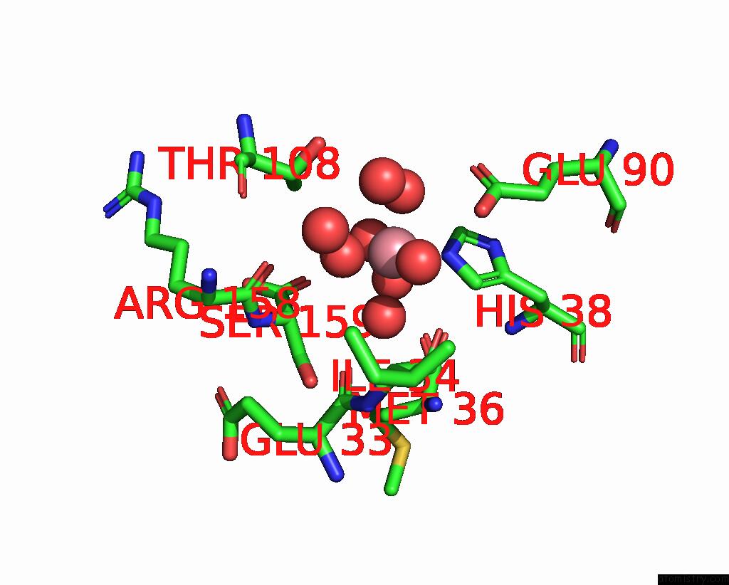
Mono view
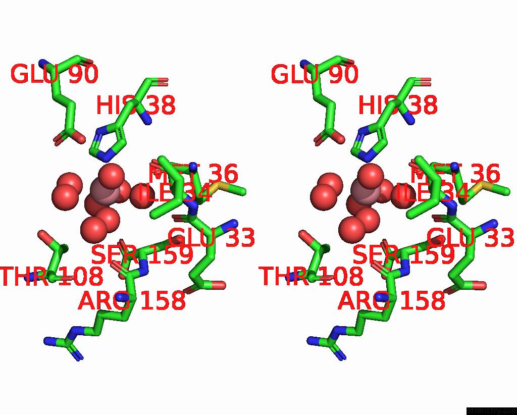
Stereo pair view
Mono view
Stereo pair view
A full contact list of Cobalt with other atoms in the Co binding
site number 2 of Structure of Dihydrofolate Reductase From Mycobacterium Tuberculosis in Complex with Nadph and Pemetrexed within 5.0Å range:
|
Reference:
J.A.Ribeiro,
S.M.Chavez-Pacheco,
G.S.De Oliveira,
C.D.S.Silva,
J.H.P.Giudice,
G.A.Libreros-Zuniga,
M.V.B.Dias.
Crystal Structures of the Closed Form of Mycobacterium Tuberculosis Dihydrofolate Reductase in Complex with Dihydrofolate and Antifolates. Acta Crystallogr D Struct V. 75 682 2019BIOL.
ISSN: ISSN 2059-7983
PubMed: 31282477
DOI: 10.1107/S205979831900901X
Page generated: Tue Jul 30 18:53:42 2024
ISSN: ISSN 2059-7983
PubMed: 31282477
DOI: 10.1107/S205979831900901X
Last articles
Zn in 9J0NZn in 9J0O
Zn in 9J0P
Zn in 9FJX
Zn in 9EKB
Zn in 9C0F
Zn in 9CAH
Zn in 9CH0
Zn in 9CH3
Zn in 9CH1