Cobalt »
PDB 6oxd-6vv9 »
6p2h »
Cobalt in PDB 6p2h: Structural Basis For 2'-Deoxyguanosine Recognition By the 2'-Dg-II Class of Riboswitches
Protein crystallography data
The structure of Structural Basis For 2'-Deoxyguanosine Recognition By the 2'-Dg-II Class of Riboswitches, PDB code: 6p2h
was solved by
M.M.Matyjasik,
R.T.Batey,
with X-Ray Crystallography technique. A brief refinement statistics is given in the table below:
| Resolution Low / High (Å) | 29.95 / 2.80 |
| Space group | P 41 2 2 |
| Cell size a, b, c (Å), α, β, γ (°) | 91.805, 91.805, 77.756, 90.00, 90.00, 90.00 |
| R / Rfree (%) | 24.4 / 27.6 |
Other elements in 6p2h:
The structure of Structural Basis For 2'-Deoxyguanosine Recognition By the 2'-Dg-II Class of Riboswitches also contains other interesting chemical elements:
| Magnesium | (Mg) | 5 atoms |
Cobalt Binding Sites:
The binding sites of Cobalt atom in the Structural Basis For 2'-Deoxyguanosine Recognition By the 2'-Dg-II Class of Riboswitches
(pdb code 6p2h). This binding sites where shown within
5.0 Angstroms radius around Cobalt atom.
In total 5 binding sites of Cobalt where determined in the Structural Basis For 2'-Deoxyguanosine Recognition By the 2'-Dg-II Class of Riboswitches, PDB code: 6p2h:
Jump to Cobalt binding site number: 1; 2; 3; 4; 5;
In total 5 binding sites of Cobalt where determined in the Structural Basis For 2'-Deoxyguanosine Recognition By the 2'-Dg-II Class of Riboswitches, PDB code: 6p2h:
Jump to Cobalt binding site number: 1; 2; 3; 4; 5;
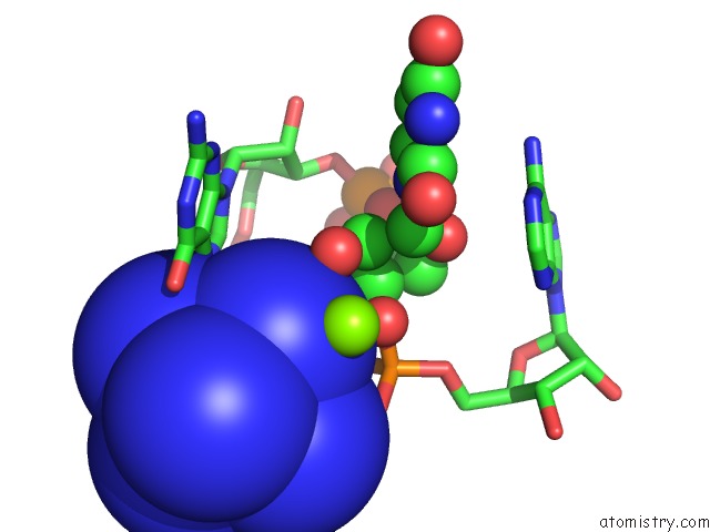
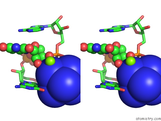
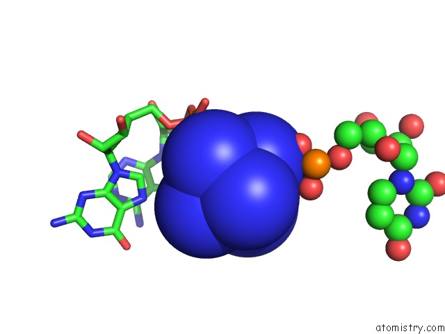
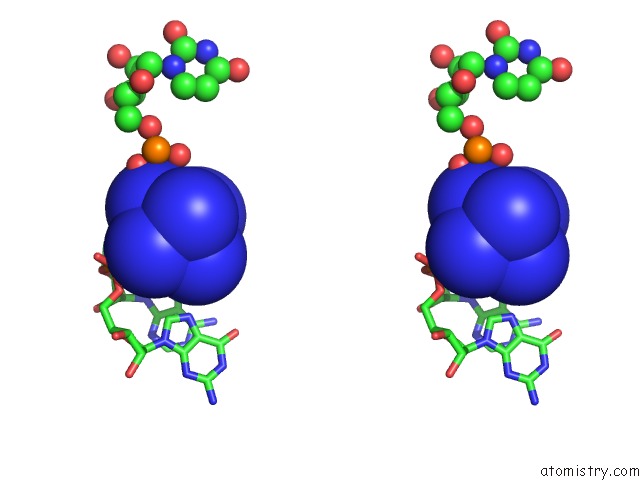
Cobalt binding site 1 out of 5 in 6p2h
Go back to
Cobalt binding site 1 out
of 5 in the Structural Basis For 2'-Deoxyguanosine Recognition By the 2'-Dg-II Class of Riboswitches
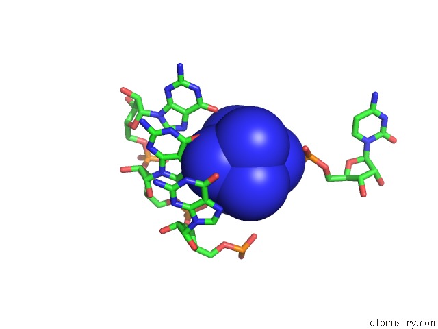
Mono view
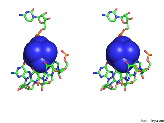
Stereo pair view
Mono view
Stereo pair view
A full contact list of Cobalt with other atoms in the Co binding
site number 1 of Structural Basis For 2'-Deoxyguanosine Recognition By the 2'-Dg-II Class of Riboswitches within 5.0Å range:
|
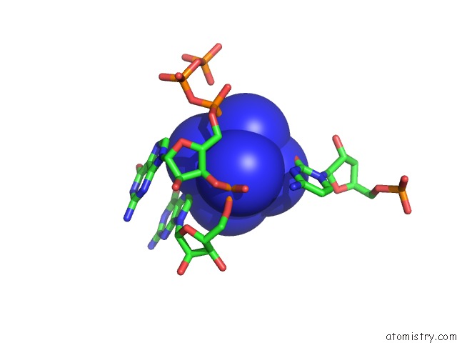
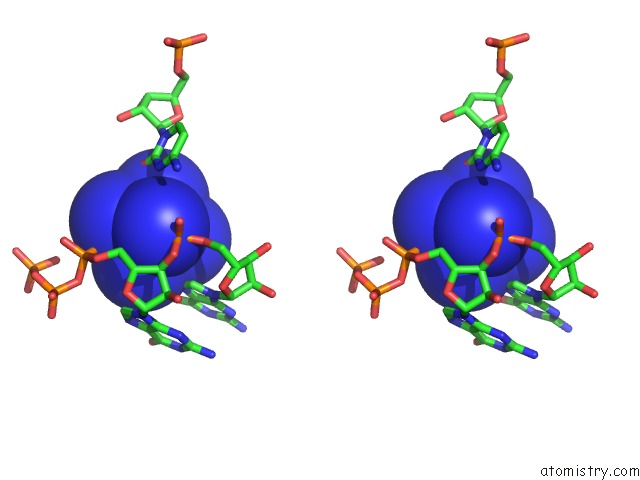
Cobalt binding site 2 out of 5 in 6p2h
Go back to
Cobalt binding site 2 out
of 5 in the Structural Basis For 2'-Deoxyguanosine Recognition By the 2'-Dg-II Class of Riboswitches
Mono view
Stereo pair view
Mono view
Stereo pair view
A full contact list of Cobalt with other atoms in the Co binding
site number 2 of Structural Basis For 2'-Deoxyguanosine Recognition By the 2'-Dg-II Class of Riboswitches within 5.0Å range:
|
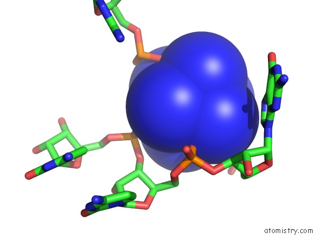
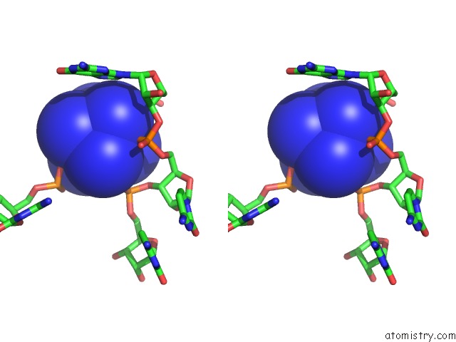
Cobalt binding site 3 out of 5 in 6p2h
Go back to
Cobalt binding site 3 out
of 5 in the Structural Basis For 2'-Deoxyguanosine Recognition By the 2'-Dg-II Class of Riboswitches
Mono view
Stereo pair view
Mono view
Stereo pair view
A full contact list of Cobalt with other atoms in the Co binding
site number 3 of Structural Basis For 2'-Deoxyguanosine Recognition By the 2'-Dg-II Class of Riboswitches within 5.0Å range:
|
Cobalt binding site 4 out of 5 in 6p2h
Go back to
Cobalt binding site 4 out
of 5 in the Structural Basis For 2'-Deoxyguanosine Recognition By the 2'-Dg-II Class of Riboswitches
Mono view
Stereo pair view
Mono view
Stereo pair view
A full contact list of Cobalt with other atoms in the Co binding
site number 4 of Structural Basis For 2'-Deoxyguanosine Recognition By the 2'-Dg-II Class of Riboswitches within 5.0Å range:
|
Cobalt binding site 5 out of 5 in 6p2h
Go back to
Cobalt binding site 5 out
of 5 in the Structural Basis For 2'-Deoxyguanosine Recognition By the 2'-Dg-II Class of Riboswitches
Mono view
Stereo pair view
Mono view
Stereo pair view
A full contact list of Cobalt with other atoms in the Co binding
site number 5 of Structural Basis For 2'-Deoxyguanosine Recognition By the 2'-Dg-II Class of Riboswitches within 5.0Å range:
|
Reference:
M.M.Matyjasik,
R.T.Batey.
Structural Basis For 2'-Deoxyguanosine Recognition By the 2'-Dg-II Class of Riboswitches. Nucleic Acids Res. V. 47 10931 2019.
ISSN: ESSN 1362-4962
PubMed: 31598729
DOI: 10.1093/NAR/GKZ839
Page generated: Tue Jul 30 18:56:43 2024
ISSN: ESSN 1362-4962
PubMed: 31598729
DOI: 10.1093/NAR/GKZ839
Last articles
Zn in 9J0NZn in 9J0O
Zn in 9J0P
Zn in 9FJX
Zn in 9EKB
Zn in 9C0F
Zn in 9CAH
Zn in 9CH0
Zn in 9CH3
Zn in 9CH1