Cobalt »
PDB 7w6v-7yvx »
7ygp »
Cobalt in PDB 7ygp: Dna Duplex Containing 5OHU-T Base Pairs
Protein crystallography data
The structure of Dna Duplex Containing 5OHU-T Base Pairs, PDB code: 7ygp
was solved by
J.Kondo,
H.Torigoe,
F.Arakawa,
with X-Ray Crystallography technique. A brief refinement statistics is given in the table below:
| Resolution Low / High (Å) | 20.79 / 2.00 |
| Space group | P 31 2 1 |
| Cell size a, b, c (Å), α, β, γ (°) | 36.672, 36.672, 54.99, 90, 90, 120 |
| R / Rfree (%) | 21.9 / 22.5 |
Cobalt Binding Sites:
The binding sites of Cobalt atom in the Dna Duplex Containing 5OHU-T Base Pairs
(pdb code 7ygp). This binding sites where shown within
5.0 Angstroms radius around Cobalt atom.
In total 2 binding sites of Cobalt where determined in the Dna Duplex Containing 5OHU-T Base Pairs, PDB code: 7ygp:
Jump to Cobalt binding site number: 1; 2;
In total 2 binding sites of Cobalt where determined in the Dna Duplex Containing 5OHU-T Base Pairs, PDB code: 7ygp:
Jump to Cobalt binding site number: 1; 2;
Cobalt binding site 1 out of 2 in 7ygp
Go back to
Cobalt binding site 1 out
of 2 in the Dna Duplex Containing 5OHU-T Base Pairs
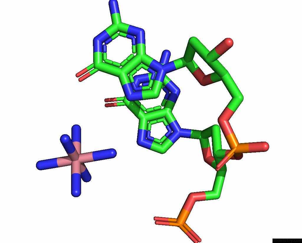
Mono view
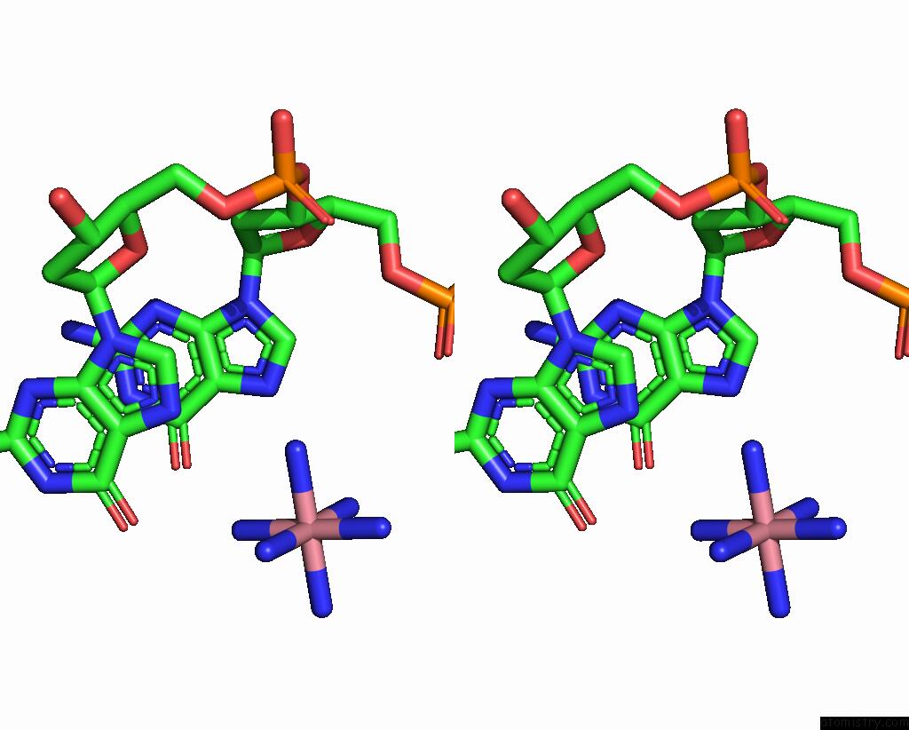
Stereo pair view
Mono view
Stereo pair view
A full contact list of Cobalt with other atoms in the Co binding
site number 1 of Dna Duplex Containing 5OHU-T Base Pairs within 5.0Å range:
|
Cobalt binding site 2 out of 2 in 7ygp
Go back to
Cobalt binding site 2 out
of 2 in the Dna Duplex Containing 5OHU-T Base Pairs
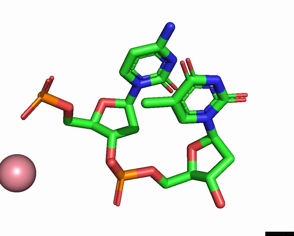
Mono view
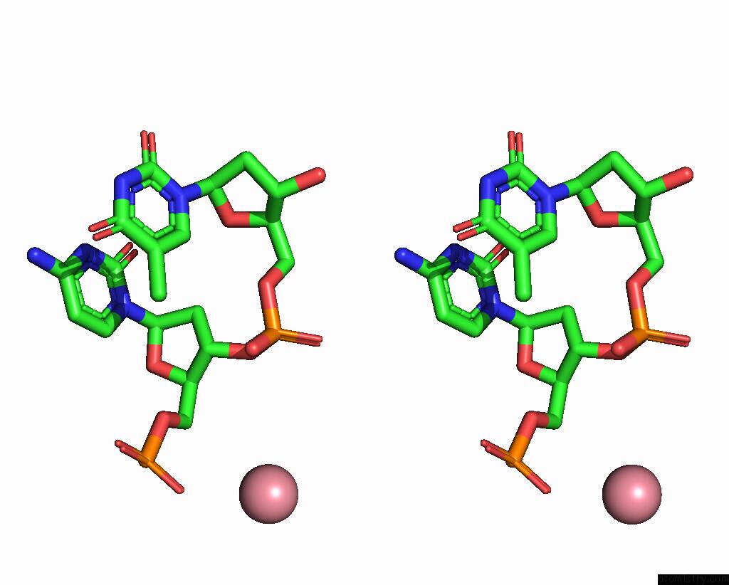
Stereo pair view
Mono view
Stereo pair view
A full contact list of Cobalt with other atoms in the Co binding
site number 2 of Dna Duplex Containing 5OHU-T Base Pairs within 5.0Å range:
|
Reference:
H.Torigoe,
J.Kondo,
F.Arakawa.
Specific Binding of Hg 2+ to Mismatched Base Pairs Involving 5-Hydroxyuracil in Duplex Dna. J.Inorg.Biochem. V. 241 12125 2023.
ISSN: ISSN 0162-0134
PubMed: 36716510
DOI: 10.1016/J.JINORGBIO.2023.112125
Page generated: Tue Jul 30 19:38:50 2024
ISSN: ISSN 0162-0134
PubMed: 36716510
DOI: 10.1016/J.JINORGBIO.2023.112125
Last articles
Zn in 9J0NZn in 9J0O
Zn in 9J0P
Zn in 9FJX
Zn in 9EKB
Zn in 9C0F
Zn in 9CAH
Zn in 9CH0
Zn in 9CH3
Zn in 9CH1