Cobalt in PDB 8bb0: The Surface-Exposed Lipo-Protein of BTUG2 in Complex with Hydroxycobalamin.
Protein crystallography data
The structure of The Surface-Exposed Lipo-Protein of BTUG2 in Complex with Hydroxycobalamin., PDB code: 8bb0
was solved by
J.Whittaker,
J.M.Felices Martinez,
A.Guskov,
D.J.Slotboom,
with X-Ray Crystallography technique. A brief refinement statistics is given in the table below:
Resolution Low / High (Å)
|
62.40 /
1.50
|
Space group
|
P 1 21 1
|
Cell size a, b, c (Å), α, β, γ (°)
|
49.11,
101.099,
79.893,
90,
97.81,
90
|
R / Rfree (%)
|
14.3 /
17.5
|
Other elements in 8bb0:
The structure of The Surface-Exposed Lipo-Protein of BTUG2 in Complex with Hydroxycobalamin. also contains other interesting chemical elements:
Cobalt Binding Sites:
The binding sites of Cobalt atom in the The Surface-Exposed Lipo-Protein of BTUG2 in Complex with Hydroxycobalamin.
(pdb code 8bb0). This binding sites where shown within
5.0 Angstroms radius around Cobalt atom.
In total 2 binding sites of Cobalt where determined in the
The Surface-Exposed Lipo-Protein of BTUG2 in Complex with Hydroxycobalamin., PDB code: 8bb0:
Jump to Cobalt binding site number:
1;
2;
Cobalt binding site 1 out
of 2 in 8bb0
Go back to
Cobalt Binding Sites List in 8bb0
Cobalt binding site 1 out
of 2 in the The Surface-Exposed Lipo-Protein of BTUG2 in Complex with Hydroxycobalamin.
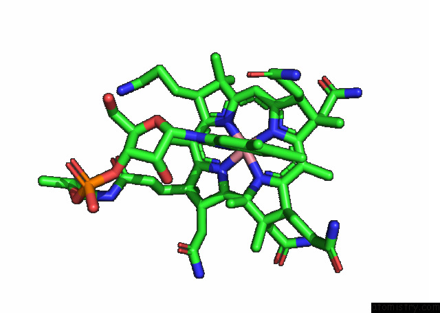
 Mono view
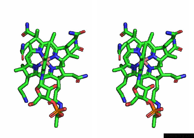
 Stereo pair view
|
A full contact list of Cobalt with other atoms in the Co binding
site number 1 of The Surface-Exposed Lipo-Protein of BTUG2 in Complex with Hydroxycobalamin. within 5.0Å range:
probe
|
atom
|
residue
|
distance (Å)
|
B
|
Occ
|
A:Co426
b:13.4
occ:1.00
|
CO
|
A:I2A426
|
0.0
|
13.4
|
1.0
|
N01
|
A:I2A426
|
1.9
|
12.2
|
1.0
|
N04
|
A:I2A426
|
1.9
|
13.4
|
1.0
|
N03
|
A:I2A426
|
1.9
|
14.3
|
1.0
|
N02
|
A:I2A426
|
1.9
|
12.9
|
1.0
|
N
|
A:I2A426
|
2.3
|
13.1
|
1.0
|
O01
|
A:I2A426
|
2.3
|
14.2
|
1.0
|
C19
|
A:I2A426
|
2.8
|
12.7
|
1.0
|
C11
|
A:I2A426
|
2.9
|
14.4
|
1.0
|
C04
|
A:I2A426
|
2.9
|
11.6
|
1.0
|
C01
|
A:I2A426
|
2.9
|
12.1
|
1.0
|
C14
|
A:I2A426
|
2.9
|
14.8
|
1.0
|
C06
|
A:I2A426
|
2.9
|
12.0
|
1.0
|
C09
|
A:I2A426
|
2.9
|
13.2
|
1.0
|
C16
|
A:I2A426
|
3.0
|
13.4
|
1.0
|
C54
|
A:I2A426
|
3.1
|
13.0
|
1.0
|
C60
|
A:I2A426
|
3.3
|
13.2
|
1.0
|
C10
|
A:I2A426
|
3.3
|
14.1
|
1.0
|
C05
|
A:I2A426
|
3.3
|
11.5
|
1.0
|
C15
|
A:I2A426
|
3.3
|
13.7
|
1.0
|
C20
|
A:I2A426
|
3.5
|
12.1
|
1.0
|
C55
|
A:I2A426
|
3.8
|
12.7
|
1.0
|
C18
|
A:I2A426
|
4.1
|
12.5
|
1.0
|
C02
|
A:I2A426
|
4.1
|
11.9
|
1.0
|
C12
|
A:I2A426
|
4.2
|
16.1
|
1.0
|
C03
|
A:I2A426
|
4.2
|
12.2
|
1.0
|
C08
|
A:I2A426
|
4.2
|
13.2
|
1.0
|
C13
|
A:I2A426
|
4.2
|
16.1
|
1.0
|
C07
|
A:I2A426
|
4.2
|
13.0
|
1.0
|
C17
|
A:I2A426
|
4.3
|
13.2
|
1.0
|
N12
|
A:I2A426
|
4.3
|
13.7
|
1.0
|
C59
|
A:I2A426
|
4.5
|
13.0
|
1.0
|
C22
|
A:I2A426
|
4.6
|
12.1
|
1.0
|
C29
|
A:I2A426
|
4.7
|
13.5
|
1.0
|
C34
|
A:I2A426
|
4.8
|
16.4
|
1.0
|
C27
|
A:I2A426
|
4.8
|
11.5
|
1.0
|
C39
|
A:I2A426
|
4.8
|
13.5
|
1.0
|
C31
|
A:I2A426
|
4.9
|
13.7
|
1.0
|
C40
|
A:I2A426
|
4.9
|
13.5
|
1.0
|
|
Cobalt binding site 2 out
of 2 in 8bb0
Go back to
Cobalt Binding Sites List in 8bb0
Cobalt binding site 2 out
of 2 in the The Surface-Exposed Lipo-Protein of BTUG2 in Complex with Hydroxycobalamin.
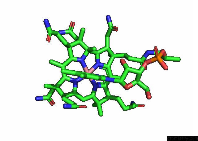
 Mono view
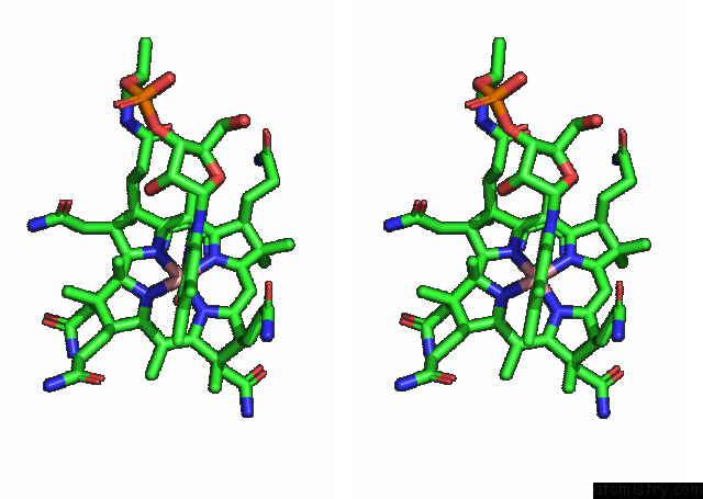
 Stereo pair view
|
A full contact list of Cobalt with other atoms in the Co binding
site number 2 of The Surface-Exposed Lipo-Protein of BTUG2 in Complex with Hydroxycobalamin. within 5.0Å range:
probe
|
atom
|
residue
|
distance (Å)
|
B
|
Occ
|
B:Co418
b:14.1
occ:1.00
|
CO
|
B:I2A418
|
0.0
|
14.1
|
1.0
|
N01
|
B:I2A418
|
1.9
|
14.0
|
1.0
|
N03
|
B:I2A418
|
1.9
|
14.3
|
1.0
|
N04
|
B:I2A418
|
1.9
|
14.9
|
1.0
|
N02
|
B:I2A418
|
1.9
|
13.3
|
1.0
|
N
|
B:I2A418
|
2.2
|
22.1
|
1.0
|
O01
|
B:I2A418
|
2.3
|
15.8
|
1.0
|
C04
|
B:I2A418
|
2.8
|
13.0
|
1.0
|
C19
|
B:I2A418
|
2.9
|
13.5
|
1.0
|
C14
|
B:I2A418
|
2.9
|
13.9
|
1.0
|
C11
|
B:I2A418
|
2.9
|
14.2
|
1.0
|
C01
|
B:I2A418
|
2.9
|
13.7
|
1.0
|
C09
|
B:I2A418
|
2.9
|
14.0
|
1.0
|
C16
|
B:I2A418
|
3.0
|
14.7
|
1.0
|
C06
|
B:I2A418
|
3.0
|
13.6
|
1.0
|
C54
|
B:I2A418
|
3.1
|
20.6
|
1.0
|
C60
|
B:I2A418
|
3.2
|
21.0
|
1.0
|
C10
|
B:I2A418
|
3.3
|
14.2
|
1.0
|
C05
|
B:I2A418
|
3.3
|
12.8
|
1.0
|
C15
|
B:I2A418
|
3.3
|
14.6
|
1.0
|
C20
|
B:I2A418
|
3.6
|
13.4
|
1.0
|
C55
|
B:I2A418
|
3.8
|
20.7
|
1.0
|
C18
|
B:I2A418
|
4.1
|
12.7
|
1.0
|
C02
|
B:I2A418
|
4.1
|
13.3
|
1.0
|
C03
|
B:I2A418
|
4.2
|
13.4
|
1.0
|
C12
|
B:I2A418
|
4.2
|
16.5
|
1.0
|
C08
|
B:I2A418
|
4.2
|
14.7
|
1.0
|
C13
|
B:I2A418
|
4.2
|
15.8
|
1.0
|
C17
|
B:I2A418
|
4.2
|
13.6
|
1.0
|
C07
|
B:I2A418
|
4.3
|
14.6
|
1.0
|
N12
|
B:I2A418
|
4.3
|
19.7
|
1.0
|
C59
|
B:I2A418
|
4.4
|
19.8
|
1.0
|
C22
|
B:I2A418
|
4.6
|
13.5
|
1.0
|
C29
|
B:I2A418
|
4.7
|
14.6
|
1.0
|
C27
|
B:I2A418
|
4.8
|
13.1
|
1.0
|
C39
|
B:I2A418
|
4.8
|
15.0
|
1.0
|
C34
|
B:I2A418
|
4.9
|
18.3
|
1.0
|
C31
|
B:I2A418
|
4.9
|
15.8
|
1.0
|
C40
|
B:I2A418
|
4.9
|
13.6
|
1.0
|
C36
|
B:I2A418
|
4.9
|
16.9
|
1.0
|
|
Reference:
J.Whittaker,
A.Guskov.
The Surface-Exposed Lipo-Protein of BTUG2 in Complex with Hydroxycobinamide. To Be Published.
Page generated: Tue Jul 30 19:42:02 2024
|